 11 citations
,
April 2016 in “The American Journal of Dermatopathology”
11 citations
,
April 2016 in “The American Journal of Dermatopathology” Special and immunohistochemical stains are not routinely needed for diagnosing hair disorders.
 5 citations
,
April 2016 in “The American Journal of Dermatopathology”
5 citations
,
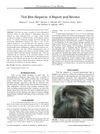
April 2016 in “The American Journal of Dermatopathology” A tick bite caused temporary hair loss in a man, which is a rare condition that usually gets better within 3 months.
 13 citations
,
March 2016 in “Journal of Cutaneous Pathology”
13 citations
,
March 2016 in “Journal of Cutaneous Pathology” Some people's hair loss is caused by multiple factors, with the most common being a mix of AGA and CCCA.
 19 citations
,
February 2016 in “Journal of The American Academy of Dermatology”
19 citations
,
February 2016 in “Journal of The American Academy of Dermatology” CD3+ T-cell presence is a reliable marker to tell apart alopecia areata from pattern hair loss.
 106 citations
,
December 2015 in “Journal of The American Academy of Dermatology”
106 citations
,
December 2015 in “Journal of The American Academy of Dermatology” Correct skin biopsy techniques are crucial to avoid misdiagnosis of skin diseases.
 31 citations
,
May 2015 in “Stem Cell Reports”
31 citations
,
May 2015 in “Stem Cell Reports” Stem cells and their surrounding environment in hair follicles work closely together, affecting hair growth and having implications for cancer and tissue regeneration.
19 citations
,
January 2015 in “Skin appendage disorders” The report found a new type of hair loss in African-American women that affects more areas of the scalp than previously thought.
 5 citations
,
January 2015 in “Current problems in dermatology”
5 citations
,
January 2015 in “Current problems in dermatology” The document concludes that a thorough history, physical exam, and specific tests are crucial for diagnosing and managing hair loss effectively.
 8 citations
,
August 2014 in “Clinical and Experimental Dermatology”
8 citations
,
August 2014 in “Clinical and Experimental Dermatology” CTE and FPHL are different hair loss types with unique causes.
 69 citations
,
August 2014 in “Journal of The American Academy of Dermatology”
69 citations
,
August 2014 in “Journal of The American Academy of Dermatology” Trichoscopy is a quick, cost-effective tool for diagnosing different hair loss conditions.
 339 citations
,
February 2014 in “Journal of The American Academy of Dermatology”
339 citations
,
February 2014 in “Journal of The American Academy of Dermatology” Most patients with frontal fibrosing alopecia are postmenopausal women, and treatments like finasteride and dutasteride can improve or stabilize the condition.
 12 citations
,
June 2013 in “The American Journal of Dermatopathology”
12 citations
,
June 2013 in “The American Journal of Dermatopathology” A new method using visual aids to diagnose hair diseases was effective after brief training.
 18 citations
,
May 2013 in “Journal of The American Academy of Dermatology”
18 citations
,
May 2013 in “Journal of The American Academy of Dermatology” EVG staining is the most reliable method for diagnosing alopecia.
 18 citations
,
October 2012 in “Dermatologic Clinics”
18 citations
,
October 2012 in “Dermatologic Clinics” Early diagnosis and aggressive treatment are key for managing rare scalp disorders that cause permanent hair loss.
 45 citations
,
May 2012 in “CRC Press eBooks”
45 citations
,
May 2012 in “CRC Press eBooks” The book helps doctors better understand and treat hair disorders due to gaps in their training.
 33 citations
,
April 2012 in “British Journal of Dermatology”
33 citations
,
April 2012 in “British Journal of Dermatology” Damaged hair follicle stem cells can cause permanent hair loss, but understanding their role could lead to new treatments.
 44 citations
,
April 2012 in “American Journal of Clinical Dermatology”
44 citations
,
April 2012 in “American Journal of Clinical Dermatology” Scarring alopecias are complex hair loss disorders that require early treatment to prevent permanent hair loss.
 8 citations
,
September 2011 in “European Journal of Dermatology”
8 citations
,
September 2011 in “European Journal of Dermatology” Most treatments for Frontal Fibrosing Alopecia are ineffective, but early anti-inflammatory therapy may help and the condition may stabilize over time.
 76 citations
,
July 2011 in “Clinical, Cosmetic and Investigational Dermatology”
76 citations
,
July 2011 in “Clinical, Cosmetic and Investigational Dermatology” The document concludes that proper diagnosis and FDA-approved treatments for different types of hair loss exist, but treatments for severe cases often fail and future improvements may focus on hair follicle stem cells.
 57 citations
,
March 2011 in “The American Journal of Dermatopathology”
57 citations
,
March 2011 in “The American Journal of Dermatopathology” Chemotherapy can cause permanent, non-reversible hair loss similar to pattern baldness.
 72 citations
,
February 2011 in “The American Journal of Dermatopathology”
72 citations
,
February 2011 in “The American Journal of Dermatopathology” Anti-TNF therapy can cause a unique type of hair loss that may get better with topical treatments without stopping the therapy.
 44 citations
,
January 2011 in “Journal of Cutaneous Pathology”
44 citations
,
January 2011 in “Journal of Cutaneous Pathology” The HoVert technique is a simple, cost-effective new method that improves alopecia diagnosis by allowing detailed analysis from a single biopsy.
 31 citations
,
December 2010 in “Journal of the American Academy of Dermatology”
31 citations
,
December 2010 in “Journal of the American Academy of Dermatology” Loose anagen hair syndrome is caused by structural abnormalities in the hair follicle's inner root sheath.
28 citations
,
June 2010 in “Pediatric dermatology” Short anagen syndrome causes short hair that may grow longer after puberty.
 10 citations
,
May 2010 in “Journal of The American Academy of Dermatology”
10 citations
,
May 2010 in “Journal of The American Academy of Dermatology” A 38-year-old African American woman has a rare condition that prevents her from growing long hair.
 67 citations
,
May 2010 in “Journal of The American Academy of Dermatology”
67 citations
,
May 2010 in “Journal of The American Academy of Dermatology” Some chemotherapy can cause permanent hair loss.
 16 citations
,
February 2010 in “Journal of the European Academy of Dermatology and Venereology”
16 citations
,
February 2010 in “Journal of the European Academy of Dermatology and Venereology” Fibrosing alopecia in a pattern distribution is a unique hair loss condition that may respond to antiandrogen therapy.
 170 citations
,
December 2009 in “Histopathology”
170 citations
,
December 2009 in “Histopathology” The conclusion is that accurate diagnosis of different types of hair loss requires good teamwork between skin doctors and lab experts.
 160 citations
,
March 2009 in “Seminars in Cutaneous Medicine and Surgery”
160 citations
,
March 2009 in “Seminars in Cutaneous Medicine and Surgery” New insights show Lichen Planopilaris is a rare, scarring hair loss condition, hard to treat, mainly affecting middle-aged women, and significantly impacts mental health.
 76 citations
,
June 2008 in “Journal of the American Academy of Dermatology”
76 citations
,
June 2008 in “Journal of the American Academy of Dermatology” The conclusion is that certain scalp tissue changes are characteristic of lichen planopilaris, with mucinous perifollicular fibroplasia being a new feature for diagnosis.
28 citations
,
June 2007 in “Journal of Cutaneous Pathology” IRS premature desquamation is not unique to CCCA and occurs in various scarring alopecias.
 100 citations
,
June 2006 in “British Journal of Dermatology”
100 citations
,
June 2006 in “British Journal of Dermatology” Hair loss severity relates to increased miniaturization in female pattern hair loss.
 126 citations
,
April 2006 in “International Journal of Dermatology”
126 citations
,
April 2006 in “International Journal of Dermatology” The conclusion is that FFA and LPP have similar scalp biopsy features, making them hard to distinguish histologically, and FFA may be a specific kind of scarring hair loss.
 33 citations
,
August 2005 in “The American Journal of Dermatopathology”
33 citations
,
August 2005 in “The American Journal of Dermatopathology” Both vertical and transverse sections are useful for diagnosing alopecia, but using both methods together is best.
 56 citations
,
July 2005 in “Journal of The American Academy of Dermatology”
56 citations
,
July 2005 in “Journal of The American Academy of Dermatology” Using both vertical and transverse sections gives a better diagnosis of alopecia than using one method alone.
 37 citations
,
July 2005 in “Journal of The American Academy of Dermatology”
37 citations
,
July 2005 in “Journal of The American Academy of Dermatology” Short anagen syndrome involves a hair growth phase lasting 1.5 years.
 179 citations
,
December 2004 in “Journal of The American Academy of Dermatology”
179 citations
,
December 2004 in “Journal of The American Academy of Dermatology” Some postmenopausal women with frontal fibrosing alopecia stopped losing hair with finasteride treatment, hinting at a possible hormonal cause.
 38 citations
,
June 2003 in “Journal of Investigative Dermatology Symposium Proceedings”
38 citations
,
June 2003 in “Journal of Investigative Dermatology Symposium Proceedings” Accurate clinical, histological, and genetic methods are key for understanding and treating hair disorders.
 30 citations
,
October 2002 in “Journal of The American Academy of Dermatology”
30 citations
,
October 2002 in “Journal of The American Academy of Dermatology” Use "female pattern hair loss" term, finasteride may help, more research needed.
 83 citations
,
January 2001 in “American journal of clinical dermatology”
83 citations
,
January 2001 in “American journal of clinical dermatology” Clomipramine may significantly reduce hair-pulling in Trichotillomania, but more research is needed on treatments and early onset cases.
 80 citations
,
March 2000 in “Journal of cutaneous pathology”
80 citations
,
March 2000 in “Journal of cutaneous pathology” The VVG stain effectively differentiates scar tissue from normal skin and helps classify types of permanent alopecia.
 158 citations
,
February 2000 in “Archives of dermatology”
158 citations
,
February 2000 in “Archives of dermatology” Some people with pattern hair loss may also have scalp inflammation and scarring similar to lichen planopilaris.
 129 citations
,
June 1999 in “Archives of Dermatology”
129 citations
,
June 1999 in “Archives of Dermatology” African Americans have less hair density than whites.
 234 citations
,
December 1996 in “Journal of The American Academy of Dermatology”
234 citations
,
December 1996 in “Journal of The American Academy of Dermatology” Middle-aged women with chronic telogen effluvium experience increased hair shedding but usually don't get significantly thinner hair.
 122 citations
,
April 1995 in “Journal of Cutaneous Pathology”
122 citations
,
April 1995 in “Journal of Cutaneous Pathology” The document describes how to tell different types of non-scarring hair loss apart by looking at hair and scalp tissue under a microscope.
 71 citations
,
March 1995 in “Journal of The American Academy of Dermatology”
71 citations
,
March 1995 in “Journal of The American Academy of Dermatology” Using both vertical and transverse sections for alopecia biopsies improves diagnosis without extra cost.
 309 citations
,
May 1993 in “Journal of The American Academy of Dermatology”
309 citations
,
May 1993 in “Journal of The American Academy of Dermatology” Horizontal scalp biopsy sections effectively diagnose and predict MPAA, with follicular density and inflammation impacting hair regrowth.
143 citations
,
October 1988 in “Clinics in Dermatology” The understanding of male-pattern baldness remains unclear.