A Histologic Review of 27 Patients with Lichen Planopilaris
June 2008
in “
Journal of the American Academy of Dermatology
”
TLDR The conclusion is that certain scalp tissue changes are characteristic of lichen planopilaris, with mucinous perifollicular fibroplasia being a new feature for diagnosis.
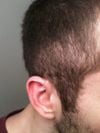
The study reviewed scalp biopsy specimens from 27 patients with confirmed lichen planopilaris (LPP) to identify key histologic features for diagnosis. The results showed that the absence of arrector pili muscles and sebaceous glands, a lymphocytic infiltrate around blood vessels and hair follicles in the reticular dermis, mucinous perifollicular fibroplasia in the upper dermis without interfollicular mucin, and superficial perifollicular wedge-shaped scarring were characteristic of LPP. The study confirmed previously reported histologic features and introduced mucinous perifollicular fibroplasia as a new diagnostic feature. However, the study was limited by the small sample size and the variability of lesions at different stages of the disease.