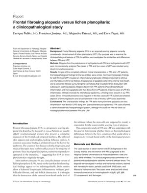
TLDR The conclusion is that FFA and LPP have similar scalp biopsy features, making them hard to distinguish histologically, and FFA may be a specific kind of scarring hair loss.
The study analyzed scalp biopsies from 16 patients, 8 with Frontal fibrosing alopecia (FFA) and 8 with lichen planopilaris (LPP), to compare their clinicopathological features. Results indicated that both conditions had similar histopathological findings, such as lymphocytic infiltrate and concentric fibrosis around hair follicles, but FFA presented with more apoptotic cells and less follicular inflammation than LPP. Additionally, FFA did not affect the interfollicular epidermis, which was sometimes involved in LPP cases. Direct immunofluorescence was negative in FFA but positive in some LPP cases. Despite these findings, the study concluded that there were no definitive histological differences to distinguish between FFA and LPP based solely on histology, and suggested that FFA might represent a specific type of lymphocytic cicatricial alopecia. However, the small sample size calls for further research to confirm these conclusions.
 57 citations
,
January 2003 in “Clinical and experimental dermatology”
 Postmenopausal frontal fibrosing alopecia is a type of hair loss in postmenopausal women that may stop on its own but has no effective treatment.
 49 citations
,
January 2003 in “Clinical and Experimental Dermatology”
 The document concludes that post-menopausal frontal fibrosing alopecia is a poorly understood condition that does not respond well to common treatments.
 89 citations
,
February 2002 in “Australasian journal of dermatology”
 A premenopausal woman had hair loss and skin issues, treated with topical steroids.
8 citations
,
January 2002 in “Piel” Postmenopausal women may experience frontal hairline and eyebrow loss due to cicatricial fibrosis.
 158 citations
,
February 2000 in “Archives of dermatology”
 Some people with pattern hair loss may also have scalp inflammation and scarring similar to lichen planopilaris.
 329 citations
,
January 1997 in “Journal of the American Academy of Dermatology”
 Frontal fibrosing alopecia is a hair loss condition in postmenopausal women, similar to lichen planopilaris, with ineffective treatments.
332 citations
,
June 1994 in “Archives of Dermatology” Postmenopausal frontal fibrosing alopecia may be a unique condition linked to postmenopausal changes.
 4 citations
,
January 2015 in “International Journal of Trichology”
 Transplanted hair follicles can change and adapt to new areas of the body, with the immune system possibly playing a role in this adjustment.
January 2014 in “Medical Innovation of China” Androgen receptor differences may contribute to hair loss in AGA patients.
 23 citations
,
November 2011 in “Journal of the European Academy of Dermatology and Venereology”
 Hair loss is a rare but recognized symptom of pemphigus vulgaris, with patients usually regrowing hair after treatment.
 12 citations
,
May 2011 in “Dermatologic Clinics”
 Hair loss in autoimmune blistering skin diseases varies and may regrow with disease control.
 51 citations
,
November 2010 in “Dermatologic Surgery”
 The research provides specific measurements for hair follicles that can improve hair transplant and regeneration techniques.
