 March 2026 in “Indian Dermatology Online Journal”
 This study reported that dermoscopy can aid in distinguishing secondary syphilis from other skin conditions by enhancing diagnostic accuracy, though there are no standardized criteria; further research is needed to confirm these findings.
3 citations
,
June 2021 in “Actas Dermo-Sifiliográficas” This study found that dermoscopy can identify typical features of Malassezia folliculitis, such as folliculocentricity and perilesional erythema, offering a valuable tool for diagnosing this condition in clinical settings.
April 2018 in “Veterinary Pathology” This study describes a form of lymphocytic mural folliculitis in tigers causing hair loss, concluding it may represent a nonspecific hypersensitivity reaction distinct from T-cell lymphoma.
9 citations
,
January 1975 in “Munich Personal RePEc Archive (Ludwig Maximilian University of Munich)” This report describes a case of Rothmund-Thomson type congenital poikiloderma, noting minor skin changes, hair loss, and slightly elevated lysine and cystine in the urine.

This study utilized polarized light microscopy to examine hair shafts in ten children with rare genetic disorders, such as Netherton syndrome and ectodermal dysplasia, providing valuable diagnostic insights into hair thickness, composition, and structural irregularities associated with these conditions.
5 citations
,
June 1993 in “Pediatric dermatology” Monilethrix Syndrome causes fragile, beaded hair that breaks easily and needs early diagnosis for better care.
 December 2014 in “Bangladesh Journal of Veterinary Medicine”
 In this study, mixed lice infestations and their associated skin changes were observed in the Egyptian lesser blind mole rat, marking the first report of such findings in this species.
 January 2024 in “Medical mycology journal”
 This study reported three cases of tinea corporis caused by Microsporum canis in a household, linked to a domestic cat, where genetic analysis suggested a familial transmission route.
16 citations
,
February 1999 in “American Journal of Dermatopathology” This article describes a woman's case of slowly progressive nonscarring alopecia with unique biopsy findings, suggesting the need to reexamine similar cases currently classified as chronic diffuse alopecia areata.
14 citations
,
August 2004 in “Veterinary Dermatology” In this study, a thoroughbred mare exhibited a unique case of alopecia areata with lymphocytic infiltration at both the bulbar and isthmic levels of hair follicles.
20 citations
,
November 2004 in “Archives of Pathology & Laboratory Medicine” This case report describes a rare instance of melanocytic matricoma, a benign pigmented papule found in sun-damaged skin of an elderly individual, highlighting its distinct clinical and pathological features.
December 2022 in “The Turkish Journal of Pediatrics” This study reported that hair microscopy can help diagnose rare pediatric neurological diseases, as specific hair characteristics were linked to conditions like giant axonal neuropathy, Griscelli syndrome, and Menkes disease.
 October 2025 in “Pediatric Dermatology”
 In this case report, a 16-year-old boy diagnosed with lupus miliaris disseminatus faciei showed improvement in facial granulomatous lesions with scarring after treatment with doxycycline, highlighting the potential need to explore treatment options for this condition characterized by asymptomatic papules on the central face.
 39 citations
,
January 2011 in “Archives of Dermatology”
39 citations
,
January 2011 in “Archives of Dermatology” This study found that the yellow dot pattern observed in dermoscopy of alopecia areata corresponds to inefficient follicular structures containing hair remnants, as corroborated by reflectance confocal microscopy and pathological findings.
March 2026 in “Mendeley Data” This abstract presents supplementary materials illustrating trichoscopy in alopecia areata and scarring alopecia using different dermoscopy techniques but reports no clinical findings.
15 citations
,
June 2020 in “Journal of the American Academy of Dermatology” Shiny white structures in trichoscopy can indicate long-standing discoid lupus erythematosus alopecia.
 2 citations
,
August 2012 in “Journal of the American Academy of Dermatology”
2 citations
,
August 2012 in “Journal of the American Academy of Dermatology” This correspondence describes two patients with both epidermolysis bullosa simplex, Dowling-Meara type, and loose anagen hair syndrome, an association not previously reported in the literature.
69 citations
,
May 2002 in “Journal of Investigative Dermatology” This study suggests that congenital atrichia with papular lesions may be more common than previously thought and proposes diagnostic criteria including the observation of hypopigmented whitish streaks on the scalp.
October 1991 in “Archives of Dermatology” This case report describes a 59-year-old woman with cicatricial alopecia and multiple skin lesions, including nodules and plaques, with biopsy results showing a lymphocytic infiltrate and collagen replacement.
53 citations
,
January 2013 in “Journal of toxicologic pathology” This publication provides a standardized nomenclature for classifying microscopic lesions in laboratory rat and mouse skin, aiming to harmonize international toxicological research.
 May 2024 in “Indian Journal of Dermatology”
 In this case report, a 22-year-old female was diagnosed with follicular Dowling-Degos disease based on clinical and histological findings, with symptoms including skin lesions confined to hair follicles. The report emphasizes the importance of differentiating this rare variant from similar conditions for proper management.
 2 citations
,
June 2012 in “Dermatologica Sinica”
2 citations
,
June 2012 in “Dermatologica Sinica” Dermoscopy is useful for diagnosing and monitoring discoid lupus erythematosus by showing specific skin patterns.
July 1979 in “Archives of Dermatology” This case report describes a 68-year-old woman with benign mucous membrane pemphigoid and unusual keratotic plaques on her extremities, associated with longstanding mouth and eye symptoms and phalangeal swelling.
 19 citations
,
April 2019 in “Journal of the European Academy of Dermatology and Venereology”
19 citations
,
April 2019 in “Journal of the European Academy of Dermatology and Venereology” This study found that acne lesion formation involves early hyperkeratinization and inflammation around hair follicles, creating a self-sustaining cycle that may progressively worsen acne.
 1 citations
,
October 2023 in “Skin research and technology”
1 citations
,
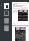
October 2023 in “Skin research and technology” This study found that line-field confocal optical coherence tomography effectively visualized key diagnostic features of classic lichen planopilaris in real time, suggesting its potential as a valuable diagnostic tool.
January 2022 in “Surgical and Cosmetic Dermatology” This case report of a 37-year-old woman with female pattern alopecia found no significant histopathological changes or follicle count improvement after three sessions of scalp microneedling.

This article discusses the clinical features of tumid lupus erythematosus and emphasizes its importance as a differential diagnosis for noncicatricial alopecia, highlighting a case similar to alopecia areata without reporting new clinical results.
2 citations
,
July 2018 in “Chinese Journal of Dermatology” This study reported that imaging characteristics of sebaceous nevus vary significantly by age group, revealing dynamic developmental changes that can be effectively identified through dermoscopy and reflectance confocal microscopy, offering noninvasive diagnostic potential.
 94 citations
,
August 1975 in “Journal of Cutaneous Pathology”
94 citations
,
August 1975 in “Journal of Cutaneous Pathology” In this study, the researchers observed that male pattern alopecia is characterized by miniature follicles, enlarged sebaceous glands, and an increased number of mast cells, without significant abnormalities in enzyme activity.
2 citations
,
June 2001 in “Medical Electron Microscopy” This study observed that the keratinization pattern of trichilemmal cysts in two Japanese women may originate from the proliferation of the outer root sheath in the follicular isthmus of anagen hairs.