Reflectance Confocal Microscopy of the Yellow Dot Pattern in Alopecia Areata
January 2011
in “
Archives of Dermatology
”
alopecia areata alopecia totalis reflectance confocal microscopy RCM yellow dot pattern dermoscopy miniaturized hair shafts broken hair shafts follicular structures empty lumina highly refractile material dilated infundibula vellus-like anagen follicles telogen follicles chronic alopecia areata alopecia hair loss yellow dots miniaturized hair broken hair hair follicles empty follicles refractile material dilated follicles vellus hair telogen hair chronic hair loss
TLDR Reflectance confocal microscopy confirms that yellow dots are signs of damaged hair follicles in alopecia areata.
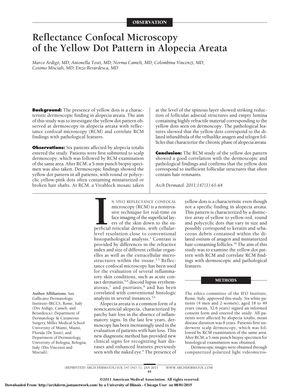
In a study from 2011, researchers used reflectance confocal microscopy (RCM) to examine the yellow dot pattern in six patients with alopecia totalis, a form of alopecia areata. Dermoscopy showed yellow dots in all patients, and these dots sometimes had miniaturized or broken hair shafts. RCM revealed a decrease in follicular structures and empty lumina with highly refractile material, which matched the yellow dots seen in dermoscopy. Pathological analysis showed that these dots were dilated infundibula of vellus-like anagen and telogen follicles, which are common in chronic alopecia areata. The study's findings demonstrated that RCM could effectively correlate with dermoscopic and pathological features, confirming that the yellow dots are indeed indicative of compromised follicular structures with occasional hair remnants.