Dermoscopic Features of Discoid Lupus Erythematosus
June 2012
in “
Dermatologica Sinica
”
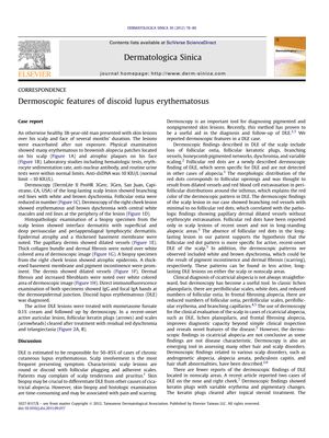
TLDR Dermoscopy is useful for diagnosing and monitoring discoid lupus erythematosus by showing specific skin patterns.
The document reports on a case of discoid lupus erythematosus (DLE) in a 38-year-old man, presenting with erythematous to brownish alopecia patches on the scalp and atrophic plaques on the face. Dermoscopic examination revealed specific features such as branching red lines, white and brown dyschromia, and reduced follicular ostia on the scalp, and erythematous and brown dyschromia with central white macules on the face. Histopathologic examination confirmed DLE, showing interface dermatitis, epidermal atrophy, thickened basement membrane, and dermal fibrosis. The patient's active DLE lesions responded to treatment with mometasone furoate cream, as observed through dermoscopy. The study highlights the utility of dermoscopy in diagnosing and monitoring DLE, noting that it can reveal disease-specific patterns, such as follicular red dots in recent-onset DLE of the scalp, and can differentiate DLE from other forms of cicatricial alopecia. Dermoscopy is emphasized as a valuable, non-invasive tool for the clinical evaluation of various hair and scalp disorders, improving diagnostic accuracy and treatment assessment.
