Dermoscopic Pattern of Cicatricial Alopecia Caused by Discoid Lupus Erythematosus and Lichen Planopilaris
April 2010
in “
Anais Brasileiros de Dermatologia
”
cicatricial alopecia discoid lupus erythematosus lichen planopilaris frontal fibrosing alopecia videodermoscopy conventional dermoscopy white patches branching capillaries keratin plugs reduced follicular ostia perifollicular scales white dots perifollicular erythema blue-grey dots interface dermatitis scarring alopecia DLE LPP FFA dermoscopy
TLDR Dermoscopy helps diagnose and monitor treatment for hair loss from scarring conditions like discoid lupus and lichen planopilaris.
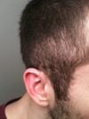
The study from 2010 investigated dermoscopic features of cicatricial alopecia in 14 patients with discoid lupus erythematosus (DLE), lichen planopilaris (LPP), and frontal fibrosing alopecia (FFA) using videodermoscopy and conventional dermoscopy. It identified distinctive dermoscopic findings for each condition: white patches, branching capillaries, keratin plugs, and reduced follicular ostia for DLE; perifollicular scales, white dots, and reduced follicular ostia for classic LPP; and reduced follicular ostia, perifollicular scales, perifollicular erythema, and branching capillaries for FFA. A novel feature, blue-grey dots, was also observed, suggesting active interface dermatitis. The study concluded that dermoscopy enhances the diagnostic process for cicatricial alopecia and can be useful for diagnosis and monitoring treatment, while also calling for larger studies to further validate these findings.
 nkrata