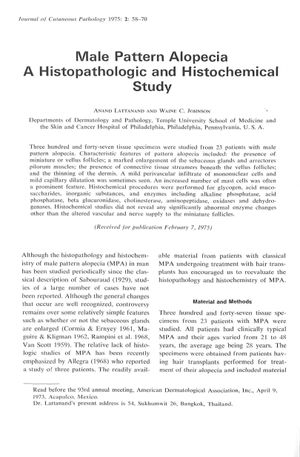
Male Pattern Alopecia: A Histopathologic and Histochemical Study
August 1975
in “
Journal of Cutaneous Pathology
”
male pattern alopecia miniature hair follicles sebaceous glands arrector pili muscles connective tissue streamers thinned dermis mast cells inflammation capillary dilatation vascular supply nerve supply anagen phase catagen-telogen follicles hyaluronic acid biopsy MPA hair follicles oil glands muscles skin tissue thin skin immune cells blood vessels nerve connections growth phase resting phase skin biopsy

TLDR Male pattern baldness involves smaller hair follicles, larger oil glands, and other tissue changes, but not major blood supply issues.
In 1975, Anand Lattanand and Waine C. Johnson conducted a study on 347 tissue specimens from 23 patients with male pattern alopecia (MPA). They discovered that MPA is characterized by miniature hair follicles, enlarged sebaceous glands and arrector pili muscles, connective tissue streamers, a thinned dermis, and an increased number of mast cells. While some specimens showed mild inflammation and capillary dilatation, histochemical studies did not indicate significant abnormal enzyme changes, except for altered vascular and nerve supply to the miniaturized follicles. The study also noted an increase in inflammatory cells in about half of the specimens, enlarged sebaceous glands, a decrease in the duration of the anagen phase, and an increase in catagen-telogen follicles. An increase in hyaluronic acid was observed in some specimens, but early MPA showed normal blood supply to follicles, suggesting that vascular insufficiency is not a cause of MPA. The study emphasized the need for adequate biopsy size for proper evaluation and differentiation from other forms of alopecia.


