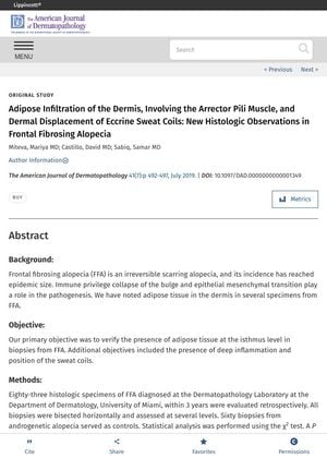
Adipose Infiltration of the Dermis, Involving the Arrector Pili Muscle, and Dermal Displacement of Eccrine Sweat Coils: New Histologic Observations in Frontal Fibrosing Alopecia
January 2019
in “
The American Journal of Dermatopathology
”
Frontal fibrosing alopecia scarring alopecia adipose tissue isthmus level deep inflammation sweat coils reticular dermis androgenetic alopecia hair follicles arrector pili muscle APM epithelial mesenchymal transition FFA scarring hair loss fat tissue sweat glands AGA hair roots muscle around hair follicle EMT
TLDR The conclusion is that fat tissue in the skin is a new finding in Frontal fibrosing alopecia and may contribute to hair follicle and muscle degeneration.
The study conducted by the Department of Dermatology and Cutaneous Surgery at the University of Miami Miller School of Medicine in 2019 examined 83 histologic specimens of Frontal fibrosing alopecia (FFA), a type of irreversible scarring alopecia. The researchers aimed to verify the presence of adipose tissue at the isthmus level in biopsies from FFA, along with the presence of deep inflammation and position of the sweat coils. Out of the 60 specimens that met the inclusion criteria, 70% showed fat tissue infiltration at the isthmus level, 55% had fat infiltration in the arrector pili muscle (APM), and 43% had sweat coils positioned in the reticular dermis. These results were significantly different from the control group of 60 biopsies from androgenetic alopecia. The study concluded that the presence of adipose tissue in the dermis is a new histologic finding in FFA, and the interaction of the hair follicles and the APM with the adipose tissue may play a role in APM degeneration and in epithelial mesenchymal transition.