 11 citations
,
May 2020 in “Anais Brasileiros de Dermatologia”
11 citations
,
May 2020 in “Anais Brasileiros de Dermatologia” This case report describes a patient with secondary syphilis exhibiting distinct dermoscopic features, including moth-eaten alopecia and an erythematous background more pronounced than previously reported.
 September 2023 in “Zenodo (CERN European Organization for Nuclear Research)”
 In this study, researchers examined trichoscopic features in 80 patients with androgenetic alopecia and found that hairshaft thickness heterogeneity was the most common feature, highlighting trichoscopy as a useful, non-invasive diagnostic tool for assessing this condition.
 12 citations
,
October 2012 in “Anais Brasileiros De Dermatologia”
12 citations
,
October 2012 in “Anais Brasileiros De Dermatologia” This study found that dermoscopy is an effective tool for diagnosing and monitoring androgenetic alopecia in women, as it helped identify common signs such as hair miniaturization and peripilar brown halo.
 40 citations
,
December 2014 in “Indian Journal of Dermatology”
40 citations
,
December 2014 in “Indian Journal of Dermatology” This study found that trichoscopy can effectively differentiate scalp psoriasis from seborrheic dermatitis by identifying specific trichoscopic patterns such as hidden hairs, signet ring vessels, and comma vessels.
 21 citations
,
January 2014 in “International Journal of Trichology”
21 citations
,
January 2014 in “International Journal of Trichology” This study observed that while dermoscopic features in alopecia areata for dark-skinned individuals align with existing literature, yellow dots appear more frequently and could help in grading disease severity.
13 citations
,
January 2018 in “Skin Appendage Disorders” This study found that dermoscopy improves clinical diagnosis accuracy for trichostasis spinulosa and proposes a new dermoscopic classification with implications for treatment outcomes.
 13 citations
,
January 2014 in “Dermatology”
13 citations
,
January 2014 in “Dermatology” This study reported that alopecic lesions after radiation exposure during angioembolization share dermoscopic features with alopecia areata, highlighting the importance of accurate patient history to avoid misdiagnosis.
 1 citations
,
October 2023 in “Veterinary Dermatology”
1 citations
,
October 2023 in “Veterinary Dermatology” This study found that in Pomeranian dogs with alopecia X, dermoscopic evaluations can supplement diagnoses, and transverse skin biopsy sections better assess hair follicle density and identify growth arrest phases compared to longitudinal sections.
 1 citations
,
October 2022 in “international journal of endocrinology and metabolism”
1 citations
,
October 2022 in “international journal of endocrinology and metabolism” This study found that patients with hypothyroidism and hair loss exhibited significant differences in dermoscopic findings, including hair shaft abnormalities and vascular changes, compared to euthyroid individuals.
This study found that dermoscopic examination can significantly distinguish between female androgenetic alopecia and telogen effluvium based on hair diameter diversity and the presence of thin and vellus hairs.
 30 citations
,
January 2014 in “Annals of Dermatology”
30 citations
,
January 2014 in “Annals of Dermatology” This study observed no significant relationship between trichoscopic findings and disease severity in male or female androgenetic alopecia, despite some specific patterns correlating with different severity levels in female patients.
4 citations
,
February 2022 in “Journal of Cosmetic Dermatology” This study found that dermatoscopic features can distinguish between tinea capitis and alopecia areata in children, with tinea capitis commonly showing comma hairs and alopecia areata showing exclamation mark hairs.
October 2023 in “Asian journal of pharmaceutical and clinical research” In this study, researchers used trichoscopy to establish standardized findings for normal hair and scalp characteristics in 360 healthy males, noting significant age-related differences in hair diameter variability and identifying potential pathological indicators like colored dots and scarring not present in healthy individuals.
 2 citations
,
August 2022 in “Clinical Case Reports”
2 citations
,
August 2022 in “Clinical Case Reports” This case report describes a 17-year-old girl with telogen effluvium and severe hair loss following a suspected COVID-19 infection and a history of similar episodes since childhood.
 May 2022 in “Authorea (Authorea)”
 This study reports a case of a 17-year-old girl experiencing telogen effluvium after COVID-19, with hair loss starting two to twelve weeks post-illness.
20 citations
,
February 2015 in “Anais brasileiros de dermatologia/Anais Brasileiros de Dermatologia” This case report describes a 6-year-old girl diagnosed with monilethrix despite no familial history and treated with a topical minoxidil trial.
7 citations
,
January 2020 in “Dermatology online journal” In this case report, adult-onset porokeratotic eccrine ostial and dermal duct nevus improved with topical tazarotene treatment, as evidenced by dermatoscopic images.
 July 2025 in “JOURNAL OF CLINICAL AND DIAGNOSTIC RESEARCH”
 In this case report, a nine-year-old boy with an unusual scalp nodule was found to have trichofolliculoma, a rare skin tumor, emphasizing the importance of considering this diagnosis in children and using dermoscopy and histopathology for accurate identification and treatment.
 February 2026 in “Clinical Cosmetic and Investigational Dermatology”
 This case report describes a 7-year-old girl who developed radiation-induced alopecia after a neurointerventional procedure, with dermoscopy revealing specific hair and scalp patterns that helped diagnose this rare complication. The findings highlight the importance of dermoscopy in distinguishing RIA from other alopecic disorders in children.
 January 2025 in “Dermatology Practical & Conceptual”
 Perinevoid alopecia can be effectively treated with non-invasive corticosteroids.
March 2026 in “Pakistan Armed Forces Medical Journal” This study found that trichoscopy positively correlates with disease severity in both male and female patients with androgenetic alopecia, suggesting its usefulness in early diagnosis and assessment.
9 citations
,
July 2020 in “JAMA dermatology” This study explored dermatoscopic and histopathologic findings of central centrifugal cicatricial alopecia beyond the vertex scalp, suggesting that dermatoscopy might serve as a less invasive diagnostic tool for subclinical disease.
 1 citations
,
November 2017 in “International Journal of Research in Medical Sciences”
1 citations
,
November 2017 in “International Journal of Research in Medical Sciences” In this study, hair follicle effacement was identified as a key dermoscopic feature of cicatricial alopecia, and dermoscopy proved useful in distinguishing cicatricial from non-cicatricial alopecias.
10 citations
,
February 2022 in “JEADV Clinical Practice” This study suggests that dermoscopy can help identify and distinguish various rosacea subtypes, improving clinical diagnosis and treatment decisions.
1 citations
,
June 2023 in “Jordan Journal of Applied Science - Natural Science Series” This study found that both topical minoxidil and latanoprost effectively improved hair regrowth in patients with alopecia areata, but minoxidil showed higher efficacy.
 6 citations
,
January 2016 in “JAMA Dermatology”
6 citations
,
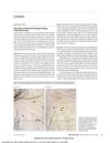
January 2016 in “JAMA Dermatology” This article discusses the presence of dirty dots as a normal trichoscopic finding in children, noting they are not observed in adults or other age groups, and reports no new results.
 January 2015 in “Indian Dermatology Online Journal”
 This diagnostic discussion reports that diffuse alopecia areata was identified in a patient with diffuse hair loss, based on specific histopathological features observed in a biopsy.
 July 2025 in “Berkala Ilmu Kesehatan Kulit dan Kelamin”
 This study described the clinical characteristics of 18 pediatric patients with alopecia areata at a hospital in Denpasar from 2021 to 2023, finding equal gender distribution and hair loss as the most common symptom, with onset typically within 1–6 months.
 January 2022 in “Clinical Cases in Dermatology”
 This review discusses the pathogenesis, diagnosis, and treatment strategies for Central Centrifugal Cicatricial Alopecia, emphasizing a multifactorial approach and reporting no new clinical results.
 12 citations
,
January 2017 in “Skin appendage disorders”
12 citations
,
January 2017 in “Skin appendage disorders” This study reports cases of histopathologically proven cutaneous lupus erythematosus manifesting as frontal fibrosing alopecia, with improvement following treatment with oral hydroxychloroquine and/or intralesional triamcinolone acetonide.