13 citations
,
September 1997 in “Archives of Dermatology” This case report describes a 5-year-old boy with a solitary patch of hair loss, potentially linked to a fungal infection after antibiotic treatment showed minimal effectiveness.
12 citations
,
April 2004 in “PubMed” This study reports the first known case of multiple type nevus lipomatosus cutaneous superficialis in a 10-month-old girl, with the unique feature of perifollicular fibrosis.
 January 2014 in “Pathology”
 Non-scarring hair loss can be diagnosed with two 4mm punch biopsies, one cut vertically and the other transversely.
 January 2011 in “Journal of The American Academy of Dermatology”
 Two patients had a rare combination of red skin spots and white scaly skin lesions not on the genitals.
June 2025 in “British Journal of Dermatology” In this report, two cases of melanocytic matricoma, a rare benign hair follicle lesion that mimics melanoma and basal cell carcinoma, were described and successfully treated with complete surgical excision. Histological analysis was crucial for differentiation from similar malignant lesions.
70 citations
,
September 2008 in “PubMed” This article reviews the role of microRNAs in skin-related conditions like psoriasis and wound healing and highlights their potential as therapeutic targets and diagnostic biomarkers, without reporting new experimental results.
 24 citations
,
September 2018 in “Lasers in Surgery and Medicine”
24 citations
,
September 2018 in “Lasers in Surgery and Medicine” This study shows that multiphoton microscopy can non-invasively distinguish between scarring and non-scarring alopecia by identifying certain morphological features, but further research is needed to confirm its clinical utility.
 100 citations
,
June 2006 in “British Journal of Dermatology”
100 citations
,
June 2006 in “British Journal of Dermatology” This study found that as hair loss severity increased in women with female pattern hair loss, the number of vellus follicles also rose, indicating miniaturization of terminal hair follicles.
 60 citations
,
September 2010 in “Journal of the American Academy of Dermatology”
60 citations
,
September 2010 in “Journal of the American Academy of Dermatology” Small white dots on the scalp seen with a dermoscope correspond to sweat ducts and vary with different hair disorders.
September 2011 in “Archives of dermatology” The child was diagnosed with cutaneous leishmaniasis.
 14 citations
,
January 2020 in “International Journal of Biological Sciences”
14 citations
,
January 2020 in “International Journal of Biological Sciences” This study explored the use of multiphoton microscopy to monitor changes in tumors and surrounding tissues in breast cancer patients undergoing neoadjuvant chemotherapy, finding it may effectively detect treatment response at cellular levels without using exogenous contrast agents.
April 2018 in “Blackwell's Five‐Minute Veterinary Consult Clinical Companion” This chapter reviews common dermatoses in small animals and offers bulleted lists as a preliminary tool for formulating differential diagnoses, but reports no new clinical findings.
66 citations
,
February 2002 in “Veterinary dermatology” This study suggests that the presence of Malassezia yeast in biopsy specimens from cats with multifocal or generalized skin lesions indicates the need for a thorough assessment for internal neoplasia.
October 2024 in “African Health Sciences” This study found that examining skin lesions with dermatoscopy aids in identifying different Traditional Chinese Medicine syndromes linked to alopecia areata.
9 citations
,
May 2013 in “European Journal of Dermatology” This article discusses eruptive vellus hair cysts, describing their clinical characteristics, and reports no new research findings.
1 citations
,
January 2021 in “Journal of the American Academy of Dermatology” This study found that bullous lupus erythematosus lesions in cutaneous lupus erythematosus showed distinctive histopathology with neutrophilic predominance and were associated with severe disease and higher extracutaneous involvement.
82 citations
,
November 1985 in “Archives of Dermatology” This case report describes a black male newborn who developed bullae that healed with hypopigmentation, linked to collagenolysis and RER vacuoles potentially containing proteolytic enzymes.
7 citations
,
February 2012 in “Journal of cutaneous pathology” This case report presents unique histopathological findings in skin lesions of hereditary mucoepithelial dysplasia that have not been previously documented.
 53 citations
,
September 2011
53 citations
,
September 2011 Other common signs, not just the well-known immune cells around hair bulbs, are important for diagnosing hair loss from alopecia areata.
3 citations
,
August 2024 in “Skin Research and Technology” In this study, LC-OCT imaging was found to noninvasively identify specific features of cicatricial alopecia due to lupus erythematosus, such as hyperreflective collagen fibers, which may aid in distinguishing it from other types of scarring alopecia.
 3 citations
,
December 2013 in “Journal of Dermatology”
3 citations
,
December 2013 in “Journal of Dermatology” This study found that scanning electron microscopy can identify four specific hair morphology patterns in alopecia areata patients, offering a less invasive diagnostic option compared to punch biopsy.
14 citations
,
August 2013 in “Journal of Parasitology” This study reported that 39.7% of Amargosa voles had mite-associated lesions, which may severely impact their health and fitness.
 May 2015 in “Actas Dermo-Sifiliográficas”
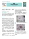
 A young man was unexpectedly diagnosed with basal cell carcinoma after a scalp examination and confocal microscopy.
June 2026 in “Indian Journal of Case Reports” This source reports a rare case of a 10-year-old child developing periorbital milia as an unusual cutaneous manifestation of chronic graft-versus-host disease following allogeneic stem cell transplantation, suggesting that immune-mediated epidermal disruption may lead to milia formation.
December 2014 in “Actas dermo-sifiliográficas/Actas dermo-sifiliográficas” Hair removal with intense pulsed light can cause rare skin lesions that are hard to fully treat.
1 citations
,
February 2023 in “American Journal of Dermatopathology” Microscarring is found in both alopecia areata and pattern hair loss, suggesting a shared cause.
 December 2022 in “The journal of pediatrics/The Journal of pediatrics”
 This case report describes a diagnosis of cutaneous pilus migrans in a 19-month-old girl, highlighting its rarity and distinction from larva migrans, and confirming it through microscopic examination.
July 2025 in “Journal of Investigative Dermatology” Scarring alopecia involves increased immune cells and specific gene changes near damaged hair follicles.
January 2023 in “Journal of Veterinary and Animal Sciences” In this study of dogs with mucocutaneous lesions caused by various dermatological conditions, the researchers observed notable changes in haemato-biochemical parameters and trichographic features, including anaemia, elevated platelet counts, and significant telogen phase hair involvement.
56 citations
,
November 1958 in “The Journal of Cell Biology” This study used electron microscopy to identify a distinct dendritic cell in the human epidermis, similar to the melanocyte, with unique structural features and variable melanin content.