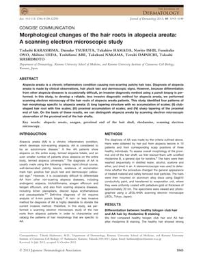
TLDR Scanning electron microscopy revealed four distinct hair root shapes in alopecia areata, suggesting a less invasive diagnostic method.
In a 2013 study, scanning electron microscopy was used to analyze the hair roots of 10 patients with alopecia areata (AA) and 3 healthy controls. The researchers discovered four unique morphological patterns in the hair roots of AA patients: long tapering structures without scale accumulation, club-shaped roots with fine scales, proximal scale accumulation, and sharp tapering at the proximal end of the hair. These patterns indicate that scanning electron microscopy could serve as a less invasive diagnostic tool for AA, providing an alternative to the more invasive punch biopsy.
35 citations
,
August 2009 in “Journal of the American Academy of Dermatology” Melanocytes might be targeted by the immune system in people with alopecia areata, but more research is needed.
 71 citations
,
March 2009 in “Seminars in cutaneous medicine and surgery”
 Alopecia areata can cause unpredictable hair loss, and treatments like corticosteroids and minoxidil may help but have varying side effects.
74 citations
,
July 2008 in “Dermatologic therapy” Early detection and histopathology are crucial to prevent permanent hair loss in cicatricial alopecia.
23 citations
,
January 2008 in “Skin Pharmacology and Physiology” Optical coherent tomography can effectively detect steroid use by analyzing hair changes.
 42 citations
,
January 2008 in “Dermatology”
 Dermoscopy effectively distinguishes between acute total hair loss and other types of female hair loss.
 44 citations
,
November 1998 in “Australasian Journal of Dermatology”
 Accurate diagnosis is key for treating different kinds of hair loss, and immune response variations may affect the condition and treatment results.
 43 citations
,
August 2013 in “Pediatric Dermatology”
 Trichoscopy is good for diagnosing and monitoring hair and scalp problems in children but needs more research for certain conditions.
 1 citations
,
April 1992 in “PubMed”
 The document describes the signs of different common types of hair loss.
31 citations
,
January 1981 PUVA-therapy is not very effective for severe hair loss types like alopecia totalis or universalis.
