Can You Diagnose Hair Loss (Non-Scarring Alopecia)
January 2014
in “
Pathology
”
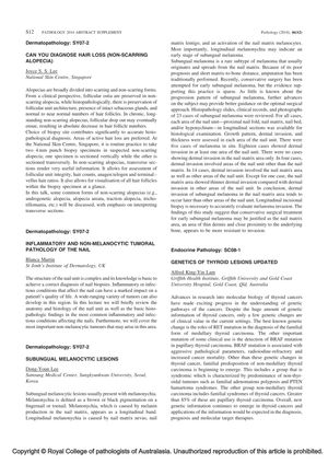
TLDR Non-scarring hair loss can be diagnosed with two 4mm punch biopsies, one cut vertically and the other transversely.
The document discusses various aspects of dermatopathology, focusing on non-scarring alopecia, nail pathology, and subungual melanocytic lesions. Joyce S. S. Lee from the National Skin Centre in Singapore highlights that non-scarring alopecias, such as androgenetic alopecia and alopecia areata, preserve follicular ostia clinically and follicular unit architecture histopathologically. Biopsies from areas of active hair loss are preferred, and two 4mm punch biopsy specimens are taken for accurate diagnosis, with one sectioned vertically and the other transversely. Transverse sections are particularly informative, allowing assessment of follicular unit integrity and hair counts. Blanca Martin and Dong-Youn Lee discuss nail pathology and subungual melanocytic lesions, respectively, with Lee reviewing 23 cases of subungual melanoma to suggest that conservative surgical treatment may be justified in early stages as the nail matrix area is more resistant to invasion. The document also briefly mentions advances in the genetics of thyroid lesions, noting the clinical importance of RET and BRAF mutations in thyroid cancers.

