Unexpected Diagnosis of Basal Cell Carcinoma
May 2015
in “
Actas Dermo-Sifiliográficas
”
basal cell carcinoma BCC melanocytic lesions dermoscopy milia-like cysts perifollicular pigmented structures in vivo reflectance confocal microscopy refractile nests nucleated cells peripheral palisading infiltrative basal cell carcinoma histological examination scalp examination hyperpigmented lesion surgical intervention skin cancer skin lesion skin examination skin surgery
TLDR A young man was unexpectedly diagnosed with basal cell carcinoma after a scalp examination and confocal microscopy.
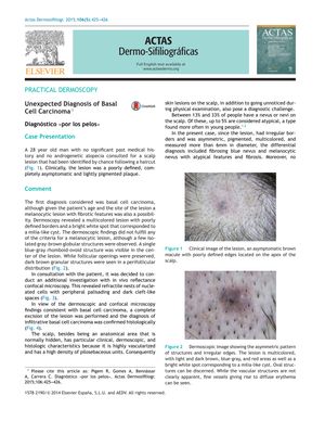
A 28-year-old man with no significant medical history presented with an asymptomatic, poorly defined, lightly pigmented plaque on his scalp, which was discovered incidentally after a haircut. Initially, basal cell carcinoma (BCC) was suspected, but melanocytic lesions were also considered due to the patient's age and lesion characteristics. Dermoscopy showed a multicolored lesion with poorly defined borders, milia-like cysts, and perifollicular pigmented structures, but did not meet the criteria for a melanocytic lesion. In vivo reflectance confocal microscopy revealed features consistent with BCC, such as refractile nests of nucleated cells with peripheral palisading. The diagnosis of infiltrative basal cell carcinoma was confirmed histologically after complete excision of the lesion. This case emphasizes the importance of thorough scalp examination, as even young patients without risk factors can develop infiltrative BCC, which may present atypically as a hyperpigmented lesion. Confocal microscopy was instrumental in confirming the diagnosis suggested by dermoscopic findings, leading to immediate surgical intervention.