 1 citations
,
May 2026 in “Signal Transduction and Targeted Therapy”
1 citations
,
May 2026 in “Signal Transduction and Targeted Therapy” This review highlights the promising role of stem cell therapy in treating resistant diseases, while also addressing the challenges in production and clinical application, emphasizing the need for standardized manufacturing and quality assurance to enhance efficacy and safety in clinical translational research.
 1 citations
,
January 2017 in “Springer eBooks”
1 citations
,
January 2017 in “Springer eBooks” This review discusses recent advances in understanding alopecia areata's disease mechanisms and highlights JAK molecules as promising therapeutic targets, but reports no new clinical results; controlled trials are needed.
 January 2026 in “Frontiers in Molecular Biosciences”
 This study identified a four-gene loop as a non-invasive biomarker that selectively activates in alopecia areata, providing a precise target for JAK inhibitor treatments.
 January 2026 in “Food Science and Human Wellness”
 This study found that oral administration of Flammulina velutipes polysaccharide reduced tumor volume and improved survival in mice by modulating macrophage polarization and enhancing antitumor immune responses.
 December 2025 in “Nature Communications”
 In this study using mouse models, researchers found that elevated IL-17a in aged olfactory epithelium impairs olfactory function, while IL-17a inhibition promotes regeneration and mitigates age-related decline.
 December 2025 in “ADMET & DMPK”
 This review synthesizes recent research to propose a precision framework for treating androgenetic alopecia and alopecia areata based on genetic insights and pathway biology, highlighting the roles of androgen-receptor signaling, immune dysregulation, and emerging therapies like regenerative medicine and AI-assisted diagnostics.
 July 2025 in “Berkala Ilmu Kesehatan Kulit dan Kelamin”
 This study described the clinical characteristics of 18 pediatric patients with alopecia areata at a hospital in Denpasar from 2021 to 2023, finding equal gender distribution and hair loss as the most common symptom, with onset typically within 1–6 months.
In this study, researchers found that CD4 T cells from the skin draining lymph nodes of mice with alopecia areata can transfer the disease to recipient mice, highlighting the key role of these cells and their interaction with CD8 T cells in the disease's development.
 January 2025 in “Repository of the Academy's Library (Library of the Hungarian Academy of Sciences)”
 In this study, baricitinib was found to be effective and safe for treating severe alopecia areata, with significant hair regrowth observed in many patients by the third month of therapy and continued improvement by the twelfth month, despite mostly mild adverse events.
 January 2025 in “Universidad de Córdoba Insitutional Repository (Universidad de Córdoba)”
 In this study, researchers observed that individuals with alopecia areata exhibited significant changes in scalp microbiota diversity and composition, which were linked to disease severity and inflammation markers, though it remains unclear if these microbial shifts are a cause or a result of hair loss.
 September 2024 in “Archives of Medical Science”
 Alopecia areata is linked to immune system differences, with specific biomarkers like CXCL9 and CXCL10 being key for diagnosis and potential treatment targets.
 June 2024 in “Research Square (Research Square)”
 This study demonstrated that in mice, the absence of Jagged-1 expression in skin-resident regulatory T cells significantly delays wound healing by reducing the accumulation of neutrophils, highlighting the role of Jagged-1 in coordinating immune cell recruitment during injury.
 December 2023 in “bioRxiv (Cold Spring Harbor Laboratory)”
 This study observed that extracellular matrix scaffold membranes performed less effectively in wound healing for aged mice compared to younger ones, with senescent SPP1+ macrophages potentially hindering epidermal and fibroblast repair abilities.
 October 2023 in “bioRxiv (Cold Spring Harbor Laboratory)”
 This study demonstrated that disrupting EGFR signaling in mice leads to immune sensitivity compromising the hair follicle's protective environment, but JAK1/2 inhibition can restore this immune privilege and stimulate hair growth, suggesting a potential therapy for scarring hair loss diseases like cicatricial alopecia.
 September 2023 in “Research Square (Research Square)”
 This study found that TNC + fibroblasts are crucial in neuro-immune interactions in various skin diseases, particularly inflammation and tumors, by engaging extensively with immune cells and overexpressing inflammatory genes, suggesting their significant role in skin abnormalities.
 February 2022 in “Research Square (Research Square)”
 This study found that high TSPEAR expression in colorectal cancer was associated with poor prognosis and correlated with various tumor and immune-related factors.
 January 2022 in “Research Square (Research Square)”
 This study found that elevated TSPEAR expression in colorectal cancer was associated with poor overall prognosis and correlated with tumor infiltrating immune cells, suggesting its potential as a predictive biomarker.
 October 2021 in “Research Square (Research Square)”
 This study found that gene expression patterns can effectively distinguish the cashmere growth cycle stages and highlight molecular pathways, suggesting melatonin's role in regulating cashmere growth in Inner Mongolian goats.
 June 2021 in “Research Square (Research Square)”
 This study reports that melatonin influences gene expression related to cashmere growth cycles in Inner Mongolian cashmere goats, potentially aiding in understanding and enhancing cashmere yield through molecular regulation.
 March 2021 in “bioRxiv (Cold Spring Harbor Laboratory)”
 This study found that type 2 immunity, especially involving ILC2 cells, helps maintain skin homeostasis by regulating the proliferation and community stability of hair follicle epithelial cells in the presence of Demodex mites.
 December 2020 in “bioRxiv (Cold Spring Harbor Laboratory)”
 In this study, 25% of CCHCR1-deficient mice exposed to stress developed hair loss similar to human alopecia areata, suggesting CCHCR1 is a susceptibility gene for the disease.
 May 2020 in “bioRxiv (Cold Spring Harbor Laboratory)”
 This study found that hair-type tissues in hedgehogs show higher enrichment of immune-related genes compared to spine-type tissues, suggesting that spines evolved to protect against injuries and infections.
 January 2023 in “Discovery immunology”
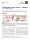
 T cells and bacteria in the gut and skin help maintain health and protect against disease.
May 2023 in “The Journal of Immunology” In this study of C3H/HeJ mice, researchers observed that alopecia areata is associated with the expansion of exhausted-memory CD8 T cells in multiple organs, with autoreactive activation not fully explained by known mechanisms, shedding light on the immunological dynamics underlying the condition.
 July 2023 in “British journal of dermatology/British journal of dermatology, Supplement”
 This study found that the E6 gene, but not E7, was responsible for HPV8-induced expansion of keratinocyte stem cells in hair follicle junctional zones in mice.
 January 2025 in “American Journal of Translational Research”
 In this study, Erianin was found to significantly suppress alopecia areata in a mouse model by inhibiting effector T cell function, reversing systemic symptoms, and promoting hair growth.
 April 2023 in “The journal of investigative dermatology/Journal of investigative dermatology”
 This study analyzed blood samples from individuals with alopecia areata and found that NKG2D+ immune cells, particularly memory-like NK cells, may better correlate with disease activity compared to CD8+ cells, though significant variability among individuals complicates these findings.
April 2026 in “Research Square”  7 citations
,
February 2015 in “Journal of comparative pathology”
7 citations
,
February 2015 in “Journal of comparative pathology” This study observed that NSG-hu-BLT mice developed graft-versus-host disease characterized by CD8+ T lymphocyte-related cell death in the skin and liver, which may affect their utility in other research areas.
This study observed two distinct groups of alopecia areata lesions based on PD-1 expression in T cells from biopsied skin samples, suggesting potential differences in subtypes or stages of the disease that warrant further investigation.