 44 citations
,
February 2015 in “Journal of the American Academy of Dermatology”
 Combining diphenylcyclopropenone with anthralin is more effective for hair regrowth in alopecia areata than using diphenylcyclopropenone alone, but may cause more side effects.
62 citations
,
January 2015 in “Journal of Dermatological Science” New genetic discoveries may lead to better treatments for alopecia areata.
52 citations
,
December 2014 in “Journal of Dermatological Science” Apremilast may help treat hair loss in alopecia areata.
701 citations
,
August 2014 in “Nature medicine” Alopecia areata can be reversed by JAK inhibitors, promoting hair regrowth.
 185 citations
,
June 2014 in “Journal of Investigative Dermatology”
 A man with severe hair loss and skin disease regrew his hair with no side effects after taking tofacitinib.
11 citations
,
December 2013 in “International Journal of Dermatology” IL16 gene variations may affect the risk of alopecia areata in Koreans.
110 citations
,
December 2013 in “The journal of investigative dermatology. Symposium proceedings/The Journal of investigative dermatology symposium proceedings” Alopecia areata is a genetic and immune-related hair loss condition that is often associated with other autoimmune diseases and does not typically cause permanent damage to hair follicles.
42 citations
,
July 2013 in “Gene” IL-4 gene variation may increase the risk of alopecia areata in Turkish people.
 22 citations
,
June 2013 in “Australasian Journal of Dermatology”
 Early stage bald spots are linked to skin inflammation and damage to the upper part of the hair follicle.
 67 citations
,
January 2013 in “Indian Journal of Dermatology, Venereology and Leprology”
 The document concludes that alopecia areata is an autoimmune disease without a definitive cure, but treatments like corticosteroids are commonly used.
19 citations
,
January 2013 in “Annals of Dermatology” Early high-dose steroid treatment helps prolong disease-free periods in severe alopecia areata.
 21 citations
,
January 2013 in “Annals of dermatology/Annals of Dermatology”
 The combination of cyclosporine and PUVA might help treat severe alopecia areata.
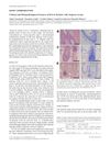 4 citations
,
January 2013 in “Acta dermato-venereologica”
 Some patients with Alopecia Areata experience itch due to immune cells and enzymes that cause itching.
 85 citations
,
October 2012 in “Dermatologic Clinics”
 Alopecia Areata is an autoimmune condition often starting before age 20, with varied treatment success and a need for personalized treatment plans.
 421 citations
,
April 2012 in “The New England Journal of Medicine”
 Alopecia Areata is an autoimmune condition causing hair loss with no cure and treatments that often don't work well.
 51 citations
,
December 2011 in “The Journal of Dermatology”
 New treatments for severe hair loss often fail, but some patients see hair regrowth with specific therapies, and treatment should be tailored to the individual's situation.
 36 citations
,
May 2011 in “Dermatologic therapy”
 No treatments fully cure or prevent alopecia areata; some help but have side effects or need more research.
 109 citations
,
May 2011 in “Dermatologic Therapy”
 Alopecia areata is a type of hair loss that can lead to complete baldness, often associated with other autoimmune conditions, and half of the cases may see hair return within a year.
 61 citations
,
September 2010 in “Genomics”
 The study found that immune responses disrupt hair growth cycles, causing hair loss in alopecia areata.
 391 citations
,
January 2010 in “Journal of The American Academy of Dermatology”
 Half of people with Alopecia Areata may see hair regrowth within a year without treatment, but recovery is unpredictable.
244 citations
,
January 2010 in “Journal of the American Academy of Dermatology” The document says current treatments for alopecia areata do not cure or prevent it, and it's hard to judge their effectiveness due to spontaneous remission and lack of studies.
36 citations
,
January 2010 in “International Journal of Trichology” Intralesional steroids can help regrow hair in some alopecia areata patients but have side effects.
138 citations
,
March 2007 in “Experimental cell research” Only a few hair-specific keratins are linked to inherited hair disorders.
 71 citations
,
August 2005 in “The journal of investigative dermatology. Symposium proceedings/The Journal of investigative dermatology symposium proceedings”
 Hair keratin-associated proteins are essential for strong hair, with over 80 genes showing specific patterns and variations among people.
 37 citations
,
November 2003 in “Veterinary pathology”
 Hair loss in mice starts with immune cells damaging hair roots before it becomes visible.
39 citations
,
April 2003 in “Australasian journal of dermatology” PUVA treatment led to significant hair regrowth in over half of the patients with alopecia areata totalis and universalis.
13 citations
,
January 2002 in “Biochemical and biophysical research communications” Interferon β from hair cells stops the growth of other hair cells.
 54 citations
,
March 1987 in “Journal of The American Academy of Dermatology”
 3% topical minoxidil effectively treats extensive alopecia areata with few side effects.
