63 citations
,
February 2003 in “Australasian Journal of Dermatology” This review discusses various methods for measuring scalp hair growth in androgenetic alopecia and reports no new results; the authors suggest that computer-assisted technology has untapped potential.
 January 2022 in “Klinicheskaya dermatologiya i venerologiya”
 This study found that anisotrichia, yellow dots, and increased vellus hairs are key trichoscopic markers of early androgenetic alopecia, while late stages are characterized by anisotrichia, yellow dots, and increased single follicular units.
41 citations
,
December 2008 in “Pediatric Dermatology” This case report indicates that trichoscopy may significantly improve the diagnosis of Netherton syndrome by noninvasively identifying typical hair abnormalities without the need to pull hair.
 1 citations
,
April 2025 in “Dermatology Practical & Conceptual”
1 citations
,
April 2025 in “Dermatology Practical & Conceptual” In this study, trichoscopy was highlighted as a valuable diagnostic tool for traction alopecia, particularly in black women, revealing common signs like vellus hair and hair casts, although it requires a suggestive clinical history for effective use.
3 citations
,
January 2017 in “Dermatology online journal” This case report describes the diagnosis of monilethrix in a 2-year-old boy using trichoscopy, highlighting its rarity and the challenge of diagnosing it without a family history.
11 citations
,
August 2016 in “Journal of the European Academy of Dermatology and Venereology” This letter discusses the use of serial trichoscopy versus a modified hair pull test for monitoring localized alopecia areata but reports no new clinical results.
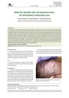 January 2026 in “Forum Dermatologicum”
 This case study examined a 72-year-old woman with unique hair shaft constrictions consistent with monilethrix, despite features atypical for this condition, ultimately reaching a diagnosis that led to successful improvement using oral minoxidil and reduced hair trauma.
18 citations
,
March 2003 in “British journal of dermatology/British journal of dermatology, Supplement” This case report details the immunohistochemical study of cytokeratins in a trichofolliculoma, providing insights but no new clinical findings.
November 2024 in “Journal of Microscopy” This study found that micro-computed tomography (micro-CT) was highly effective for analyzing the cross-sectional geometry of hair fibres, including the medulla in highly curled hair, but less effective than scanning electron microscopy for surface details.
October 2024 in “Dermatology Practical & Conceptual” This study found that trichoscopy is a valid and sensitive tool for diagnosing and evaluating eyebrow involvement in lichen planopilaris and frontal fibrosing alopecia.
1 citations
,
January 2021 in “Springer Proceedings in Materials” This study demonstrates a sample preparation method for wool follicles that allows for clear keratin labeling and ultrastructural preservation in microscopy, reducing common artefacts from conventional fixation techniques.
August 2003 in “Special report” This report introduces Barcelona - TrichoScan as a novel software tool purported to effectively analyze hair growth in patients with androgenetic alopecia or androgenetic effluvium, but it provides no clinical data.
 November 2023 in “International Journal of Dermatology”
 This review presents European expert guidelines for diagnosing androgenetic alopecia across genders but reports no new clinical findings; it emphasizes the importance of trichoscopy for differentiating similar conditions.
 June 2026 in “IP Indian Journal of Clinical and Experimental Dermatology”
 This study found that trichoscopic parameters in male androgenetic alopecia deteriorated progressively with increasing disease severity and were significantly linked to genetic, metabolic, and psychosocial factors.
 September 2025 in “Egyptian Journal of Dermatology and Venerology”
 This case study described the diagnosis of a trichofolliculoma in a 56-year-old woman, emphasizing its dermoscopic features and histological aspects that confirmed the tumor type, with no recurrence observed after excision.
 January 2023 in “Al-Azhar International Medical Journal /Al-Azhar International Medical Journal”
 Panoramic Trichoscopy is a new, better way to assess widespread hair loss.
18 citations
,
August 2009 in “Skin Research and Technology” In this study, optical coherence tomography was used to produce 3D images of human hair, revealing that after chemical treatment with ammonium thioglycolate, the main hair structures were not identifiable due to damage.
 December 2024 in “Journal of Skin and Stem Cell”
 This review discusses the role of trichoscopy in diagnosing and managing various alopecia types, highlighting its ability to detect specific patterns in both scarring and non-scarring forms, thus aiding in treatment decisions and reducing biopsy necessity.
 13 citations
,
August 2017 in “International Journal of Dermatology”
13 citations
,
August 2017 in “International Journal of Dermatology” This study reported that trichoscopy revealed distinctive patterns across different autoimmune bullous diseases, suggesting it can be a useful preliminary diagnostic tool for differentiating these conditions.
 16 citations
,
January 2011 in “International Journal of Trichology”
16 citations
,
January 2011 in “International Journal of Trichology” This article discusses the principles and considerations of clinical photography for hair disorders in dermatology, reporting no new clinical findings.
45 citations
,
December 2004 in “Forensic Science International” This study reports that using laser microdissection to isolate telogen hair follicles significantly improves DNA extraction efficiency for STR typing, reducing contamination from keratin.
 May 2026 in “Journal of Basic and Clinical Health Sciences”
 This study found that certain trichoscopic features, including hair diameter diversity patterns, helped differentiate female androgenetic alopecia from telogen effluvium in women, suggesting their potential use in clinical diagnosis.
 15 citations
,
February 2003 in “British Journal of Dermatology”
15 citations
,
February 2003 in “British Journal of Dermatology” This study demonstrated that using morphometric and 3D reconstruction software to evaluate scalp biopsies in non-cicatricial alopecias may be feasible and sensitive for detecting subtle follicular changes.
 18 citations
,
July 2018 in “Skin appendage disorders”
18 citations
,
July 2018 in “Skin appendage disorders” This review discusses trichoscopic features of hair disorders in individuals with dark skin phototypes and reports no new clinical results; the authors emphasize the need for more specific diagnostic criteria for these populations.
 September 2014 in “Proceedings of SPIE, the International Society for Optical Engineering/Proceedings of SPIE”
 This study found that high-speed optical coherence tomography may be a promising non-invasive tool for analyzing human hair conditions.
 May 2026 in “International Journal of Versatile Research and Analysis”
 This study used air wedge interferometry to measure the thickness of human scalp hair from three individuals, finding average thicknesses of 66.482 µm, 59.355 µm, and 59.927 µm respectively, within the typical range for adults.
 1 citations
,
December 2024 in “Dermatology and Therapy”
1 citations
,
December 2024 in “Dermatology and Therapy” This study found that the STRIAA scoring tool provides a rapid and highly reliable assessment of alopecia areata severity, showing a significant correlation with the traditional SALT score, thus enhancing clinical evaluation in patients with this condition.
 28 citations
,
October 1992 in “JEADV. Journal of the European Academy of Dermatology and Venereology/Journal of the European Academy of Dermatology and Venereology”
28 citations
,
October 1992 in “JEADV. Journal of the European Academy of Dermatology and Venereology/Journal of the European Academy of Dermatology and Venereology” In this study, Scalp Immersion Proxigraphy (SIP) was shown to produce more accurate measurements of linear hair growth rate than crude phototrichogram techniques, suggesting SIP's suitability for clinical trials monitoring hair growth.
37 citations
,
April 1977 in “Journal of Cutaneous Pathology” This study found that specific histologic features in skin biopsies can help support a diagnosis of traumatic alopecia (trichotillomania), but not all markers are distinct to the condition.
January 2023 in “Journal of Clinical and Diagnostic Research” This study in Chennai, India, found that while some trichoscopic patterns are specific, many overlap across different scalp dermatoses, highlighting the importance of dermatologists staying informed about the latest findings.