Histopathologic Evaluation of Alopecias
June 2006
in “The American Journal of Dermatopathology”
TLDR The document concludes that accurate diagnosis of different types of hair loss requires careful examination of hair and scalp tissue, considering both clinical and microscopic features.
The document from 2006 provides a comprehensive review of the histopathologic evaluation of alopecias, emphasizing the importance of clinicopathologic correlation for accurate diagnosis due to the overlap in histopathologic features among different types of alopecia. It describes the hair growth cycle, with anagen being the most affected phase by alopecia, and discusses the ambiguity in classifying alopecias as scarring or nonscarring, noting the prognostic importance of identifying true scarring. Elastic tissue stains are highlighted as useful in differentiating these alopecias. Androgenetic alopecia, the most common form of hair loss, is characterized by follicle miniaturization and a decreased anagen:telogen ratio, with the significance of perifollicular inflammation remaining unclear. The document also covers telogen effluvium, chronic telogen effluvium, alopecia areata, trichotillomania, and traction alopecia, detailing their histopathologic features and the necessity of histopathology for differential diagnosis. Additionally, it discusses the classification and pathogenesis of primary scarring alopecias, including stem cell failure, sebaceous gland damage, and destruction of the outer root sheath. The document further examines the differentiation between DLE, lichen planopilaris, and classic pseudopelade of Brocq, highlighting the role of special stains and immunofluorescence. Lastly, it touches on mixed inflammatory primary scarring alopecias, such as folliculitis keloidalis, and the importance of histopathologic differential diagnosis.
View this study on journals.lww.com →
Cited in this study
research Post-menopausal frontal fibrosing alopecia
The document concludes that post-menopausal frontal fibrosing alopecia is a poorly understood condition that does not respond well to common treatments.

research Postmenopausal frontal fibrosing alopecia
Postmenopausal frontal fibrosing alopecia is a type of hair loss in postmenopausal women that may stop on its own but has no effective treatment.
research Approach to the adult female patient with diffuse nonscarring alopecia
The document concludes that careful evaluation is key to diagnose and treat women with hair loss, with tests for thyroid, iron, and hormones as needed.

research Telogen effluvium
Telogen effluvium is a common type of hair loss that can resolve on its own or become chronic, with treatment depending on early diagnosis.
research Frontal fibrosing alopecia associated with cutaneous lichen planus in a premenopausal woman
A premenopausal woman had hair loss and skin issues, treated with topical steroids.
research Possible mechanisms of miniaturization during androgenetic alopecia or pattern hair loss
Hair loss occurs due to fewer papillary cells, smaller follicles, and shorter growth phases.

research Cicatricial (scarring) alopecia
Different types of scarring alopecia may be stages of one disease, and accurate diagnosis is crucial to prevent permanent hair loss.
research Lymphocytic mediated alopecia: histological classification by pattern analysis
Steven Kossard classified lymphocyte-related hair loss into four patterns, each linked to different types of baldness.
research Asebia-2J (Scd1ab2J): A New Allele and a Model for Scarring Alopecia
research Elastic tissue in scars and alopecia
research Measuring Reversal of Hair Miniaturization in Androgenetic Alopecia by Follicular Counts in Horizontal Sections of Serial Scalp Biopsies: Results of Finasteride 1mg Treatment of Men and Postmenopausal Women
Finasteride 1mg helps reverse hair miniaturization in men and postmenopausal women.
research Dermatopathology of Common Hair Problems
Examining scalp biopsies in different ways helps better diagnose hair loss types.

research The Biology of Hair Follicles
Hair follicle biology advancements may lead to better hair growth disorder treatments.
research Hair Follicle Biology, the Sebaceous Gland, and Scarring Alopecias

research Diffuse hair loss
Hair loss that spreads out can often fix itself or be treated by finding and handling the cause.

research Androgenetic alopecia
Hair loss from genetics and hormones can be treated with drugs or surgery.
research Traumatic alopecia
research Acquired scalp alopecia. Part I: A review
Accurate diagnosis is key for treating different kinds of hair loss, and immune response variations may affect the condition and treatment results.
research Eosinophils in fibrous tracts and near hair bulbs: A helpful diagnostic feature of alopecia areata
Finding eosinophils near hair bulbs helps diagnose alopecia areata.
research Postmenopausal frontal fibrosing alopecia: A frontal variant of lichen planopilaris
Frontal fibrosing alopecia is a hair loss condition in postmenopausal women, similar to lichen planopilaris, with ineffective treatments.
research Chronic telogen effluvium: Increased scalp hair shedding in middle-aged women
Middle-aged women with chronic telogen effluvium experience increased hair shedding but usually don't get significantly thinner hair.
research CHRONIC TELOGEN EFFLUVIUM
Chronic Telogen Effluvium is a hair loss condition in middle-aged women that usually doesn't lead to complete baldness.
research Histopathology of non-scarring alopecia
The document describes how to tell different types of non-scarring hair loss apart by looking at hair and scalp tissue under a microscope.
research Vertical and transverse sections of alopecia biopsy specimens: Combining the two to maximize diagnostic yield
Using both vertical and transverse sections for alopecia biopsies improves diagnosis without extra cost.

research Clinical Findings, Cutaneous Pathology, and Response to Therapy in 21 Patients With Keratosis Pilaris Atrophicans
The study found that Keratosis Pilaris Atrophicans is a genetic skin condition that starts in childhood, involves inflammation and scarring, and current treatments are only somewhat effective.
research Diagnostic and predictive value of horizontal sections of scalp biopsy specimens in male pattern androgenetic alopecia
Horizontal scalp biopsy sections effectively diagnose and predict MPAA, with follicular density and inflammation impacting hair regrowth.
research Telogen Effluvium
Telogen effluvium is a reversible hair loss condition that requires a detailed diagnosis and often resolves on its own.
research Cutaneous immunopathology of androgenetic alopecia
research Hair anatomy for the clinician
Understanding hair follicle anatomy helps diagnose hair disorders.
research Acne Keloidalis: A Review
research Histologic Response to Topically Applied Minoxidil in Male-Pattern Alopecia
Minoxidil can help grow hair and make hair follicles bigger, but it can also cause side effects.
research Androgenetic alopecia, trichotrophic substances, and histologic studies of the human scalp
Minoxidil promotes hair growth but exact mechanism is unknown.
research Androgenic Alopecia
The document concludes that treating female hair loss should target reducing excess androgen and blocking its effects on hair follicles, with the best treatments being hormonal therapy, adrenal suppression, and topical minoxidil.
research Keratosis Follicularis Spinulosa Decalvans
KFSD is a rare condition causing scarring hair loss, with no effective treatment known at the time of the report.
research Alopecia: A Pathologist's View
The review concludes that accurate diagnosis of different types of hair loss requires proper biopsy techniques and understanding the hair growth cycle and underlying causes.
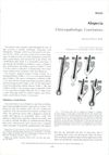
research Alopecia.
The document concludes that correct diagnosis of alopecia types is crucial, scalp biopsies are important, and more research is needed.
research Male Pattern Alopecia A Histopathologic and Histochemical Study
Male pattern baldness involves smaller hair follicles, larger oil glands, and other tissue changes, but not major blood supply issues.