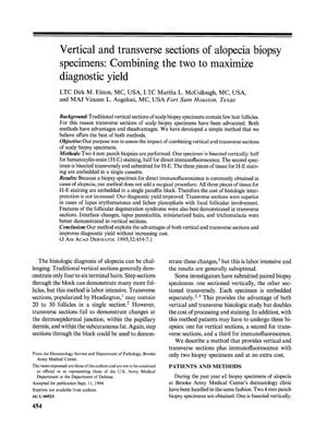
Vertical and transverse sections of alopecia biopsy specimens: Combining the two to maximize diagnostic yield
March 1995
in “
Journal of The American Academy of Dermatology
”

TLDR Using both vertical and transverse sections for alopecia biopsies improves diagnosis without extra cost.
In the 1995 study, the authors developed a method to improve the diagnostic yield of alopecia biopsy specimens by combining vertical and transverse sections without increasing costs. They performed two 4 mm punch biopsies on patients, bisecting one vertically for hematoxylin-eosin (H-E) staining and direct immunofluorescence, and the other transversely for H-E staining, embedding all three pieces in a single cassette. This method did not add a surgical procedure since a biopsy for direct immunofluorescence is common in alopecia cases. The results showed that transverse sections were superior in diagnosing lupus erythematosus and lichen planopilaris with focal follicular involvement, as well as features of the follicular degeneration syndrome. Vertical sections were better for demonstrating interface changes, lupus panniculitis, miniaturized hairs, and trichomalacia. The combined approach increased diagnostic yield, decreased laboratory costs by reducing the need for deeper levels, and was beneficial for teaching and publication purposes. The study concluded that exploiting the advantages of both sectioning methods improved diagnostic outcomes without additional costs.


