5 citations
,
November 2023 in “Journal of Investigative Dermatology” This study observed that cutaneous microwounds created by fractional photothermolysis heal differently from typical wounds, displaying rapid re-epithelialization and limited inflammation, which may help reduce scar formation by delaying collagen remodeling and minimizing myofibroblast activation.
2 citations
,
January 2022 in “Actas Dermo-Sifiliográficas” In this study, researchers found that dermoscopy revealed specific features in Malassezia folliculitis, such as folliculocentricity and perilesional erythema, which can aid in its identification and potentially distinguish it from acne vulgaris.
 February 2024 in “Skin research and technology”
 This study found that line-field confocal optical coherence tomography effectively visualized microscopic alterations in alopecia areata, suggesting its potential use in diagnosis and monitoring.
2 citations
,
May 2014 in “PubMed” This case report describes a 10-year-old boy with a localized patch of partial alopecia and comedonal lesions, diagnosed as an atypical nevus comedonicus, and treated with topical tretinoin.
March 2022 in “Veterinary dermatology” This case report provides the first evidence of clinical and histopathological findings of cutaneous diffuse basaloid follicular hamartomas in a young domestic short hair cat.
 January 2016 in “Türkiye klinikleri tıp bilimleri dergisi”
 About 38% of patients at a dermatology clinic had oral lesions, with coated tongue, fissured tongue, and Fordyce granules being most common.
September 2017 in “PubMed” In this case report, a Danish family with monilethrix showed varying symptoms, diagnosed via dermatoscopy, microscopy, and gene sequencing. The study highlights that while no cure exists, oral minoxidil shows promise in a single case, and reducing hair trauma remains key for management.
February 2023 in “Journal of the European Academy of Dermatology and Venereology” 43 citations
,
April 1996 in “Journal of Investigative Dermatology” This pilot study employed in-vivo multiphoton microscopy to visualize pigment-producing melanocytes in vitiligo patients, aiming to enhance understanding of treatment impacts and potentially improve transplantation therapies.
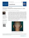 January 2026 in “Cosmoderma”
 In this clinical case study, a 9-year-old girl was diagnosed with monilethrix, a hereditary hair shaft disorder characterized by weak, beaded hair, with management focusing on minimizing hair trauma.
 September 2017 in “Journal of Investigative Dermatology”
 This study found that certain circulating miRNAs in skin and plasma may serve as potential biomarkers for distinguishing frontal fibrosing alopecia from control cases, highlighting the need for further validation in larger cohorts.
 April 2024 in “Oral Surgery Oral Medicine Oral Pathology and Oral Radiology”
 In this case report, the patient was diagnosed with oral lichen sclerosus following a detailed clinical and histological examination and referral to dermatology, and it underscores the importance of interdisciplinary collaboration in managing this rare condition and its associated risks.
 December 2025 in “Clinical Cosmetic and Investigational Dermatology”
 This case study describes a rare instance of subacute cutaneous lupus erythematosus on the chin, highlighting the effectiveness of using dermoscopy and reflectance confocal microscopy for accurate diagnosis and monitoring, which led to significant lesion improvement with corticosteroid and immunomodulatory treatment over 19 weeks.
January 2009 in “Nova Science Publishers (Nova Science Publishers, Inc.)” This study observed that human skin exhibits acute and chronic histological changes due to tick bites, including inflammation and changes in hair follicles, influenced by tick saliva and possible infections.
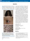 6 citations
,
August 2012 in “The Journal of Pediatrics”
6 citations
,
August 2012 in “The Journal of Pediatrics” This case report describes a 12-year-old girl diagnosed with monilethrix, characterized by fragile, beaded hair shafts, with no effective treatment currently available.
January 2009 in “Repositório Científico do Instituto Politécnico de Viseu (Instituto Politécnico de Viseu)” This case study found that a skin lesion in a Samoyed dog did not match any single known follicular cyst or neoplasm type described in the literature.
 39 citations
,
December 2008 in “Clinics in Dermatology”
39 citations
,
December 2008 in “Clinics in Dermatology” This article reviews recent trends and developments in dermoscopy, highlighting its expanding use beyond pigmented lesions, and reports no new clinical results.
37 citations
,
April 1977 in “Journal of Cutaneous Pathology” This study found that specific histologic features in skin biopsies can help support a diagnosis of traumatic alopecia (trichotillomania), but not all markers are distinct to the condition.
January 1999 in “Journal of Investigative Dermatology”  January 2022 in “Nasza Dermatologia Online”
 In this study, dermoscopy was suggested as a potentially useful tool for improving the clinical recognition of pilomatricoma.
1 citations
,
September 2022 in “European Journal of Dermatology” This study identified a novel splice-site variant of the LAMB3 gene that may cause junctional epidermolysis bullosa, suggesting gene sequencing is essential for accurate diagnosis.
 30 citations
,
October 2013 in “Lupus”
30 citations
,
October 2013 in “Lupus” This study identified distinct clinical and dermatoscopic features to differentiate non-scarring patchy alopecia in SLE from alopecia areata, emphasizing the value of serological tests in diagnosis.
September 2023 in “JEADV Clinical Practice” This study identified key dermoscopic features in folliculotropic mycosis fungoides patients, showing that dilation of follicular openings and spiky follicular keratosis are specific indicators of follicular involvement, potentially aiding in diagnosis, classification, and treatment decisions.
 November 2023 in “Berkala Ilmu Kesehatan Kulit dan Kelamin/Berkala ilmu kesehatan kulit dan kelamin (Periodical of dermatology and venerology)”
 This study suggests that using a dermoscope, a handheld microscope, can effectively help distinguish between Pityrosporum folliculitis and Acne vulgaris by revealing unique lesion characteristics that are not visible to the naked eye, offering a practical diagnostic alternative when KOH examinations are unavailable.
 10 citations
,
July 2008 in “Journal of Cutaneous Pathology”
10 citations
,
July 2008 in “Journal of Cutaneous Pathology” This study reports that marked, deep dermal mucin deposition in a patient with alopecia areata may indicate underlying systemic lupus erythematosus, as this feature was not prevalent in other alopecia areata cases.
March 2026 in “Mendeley Data” This abstract presents supplementary trichoscopy images for alopecia areata and scarring alopecia but reports no clinical findings.
33 citations
,
December 1982 in “Developmental Medicine & Child Neurology” The authors reviewed cases of six children with both hair-shaft abnormalities and neurological disorders, noting that such hair defects may indicate neurological conditions, including potentially treatable metabolic errors.
This case report describes an ulcerated lesion in the oral cavity of an 85-year-old man that has not responded to various treatments and remains without a conclusive diagnosis.
8 citations
,
July 2017 in “The journal of investigative dermatology/Journal of investigative dermatology” This study identified four circulating microRNAs as highly predictive of frontal fibrosing alopecia status, suggesting potential roles in diagnostics and disease understanding.