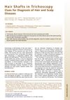
Hair Microscopy: An Easy Adjunct to Diagnosis of Systemic Diseases in Children
November 2021
in “
Applied Microscopy
”
hair microscopy light microscopy polarized light microscopy electron microscopy atomic force microscopy confocal scanning microscopy Comel-Netherton syndrome Griscelli syndrome Chediak Higashi syndrome Menkes disease Trichothiodystrophy Uncombable Hair Syndrome melanin granules sulfur-deficient hair fragile hair oral biotin supplementation biotin
TLDR Hair microscopy is a simple and cost-effective method to help diagnose systemic diseases in children.
Hair microscopy is a valuable, yet underutilized, diagnostic tool for identifying systemic diseases in children, particularly in resource-limited settings. The technique can reveal changes in hair structure and growth patterns indicative of various diseases, such as Comel-Netherton syndrome, Griscelli syndrome, Chediak Higashi syndrome, and Menkes disease. The document discusses several microscopy techniques, including light microscopy, polarized light microscopy, electron microscopy, atomic force microscopy, and confocal scanning microscopy. It also highlights specific hair features that are characteristic of certain diseases, such as the irregular distribution of melanin granules in Griscelli syndrome type 2 and regularly irregular deposition of melanin granules in Chediak Higashi syndrome. Other conditions discussed include Trichothiodystrophy, characterized by sulfur-deficient fragile hair, and Uncombable Hair Syndrome, which can be improved with oral biotin supplementation. Despite the availability of more advanced methods, light microscopy remains a useful, inexpensive, and easily accessible technique.
 Henryk