January 2016 in “Dermatology online journal” This case report describes an adolescent female with a linear array of trichoepitheliomas on her left neck, highlighting the clinical presentation of this benign tumor.
 January 2015 in “Indian Dermatology Online Journal”
 This diagnostic discussion reports that diffuse alopecia areata was identified in a patient with diffuse hair loss, based on specific histopathological features observed in a biopsy.
February 2013 in “Journal of the American Academy of Dermatology” Follicular red dots can appear where alopecia areata and vitiligo overlap.
 April 2012 in “Informa Healthcare eBooks”
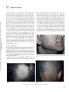
 Alopecia areata is a common autoimmune condition causing varying hair loss, diagnosed by specific patterns of inflammation around hair follicles, with several treatment options available.
 January 2012 in “Yearbook of Dermatology and Dermatologic Surgery”
 The study concluded that a 'Swiss cheese' pattern in hair follicles is a useful sign for diagnosing alopecia areata.
 January 2012 in “Yearbook of Dermatology and Dermatologic Surgery”
 Alopecia areata is a hair loss condition that often starts before age 30 and can affect various body parts, with unpredictable hair regrowth chances.
 February 2011 in “Journal of the American Academy of Dermatology”
 This study found that polycystic ovary syndrome was the most common cause of hirsutism among premenopausal women in Algeria.

The ProScope HR is an effective, user-friendly, and affordable tool for diagnosing hair loss.
April 2023 in “Dohuk medical journal” This review discusses the use of trichoscopy for diagnosing alopecia and reports no new clinical results, emphasizing its role in identifying vascular and follicular features.
November 2022 in “Al-Azhar International Medical Journal (Print)” This study observed that combining 5% topical spironolactone with 5% topical minoxidil in a single dose could enhance treatment efficacy for androgenetic alopecia in both men and women, with significant hair growth improvements noted in group C after three months.
This study found that dermoscopic examination can significantly distinguish between female androgenetic alopecia and telogen effluvium based on hair diameter diversity and the presence of thin and vellus hairs.
 January 2022 in “Klinicheskaya dermatologiya i venerologiya”
 This study found that anisotrichia, yellow dots, and increased vellus hairs are key trichoscopic markers of early androgenetic alopecia, while late stages are characterized by anisotrichia, yellow dots, and increased single follicular units.
January 2022 in “مجلة جامعة المجمعه للعلوم الصحية” This case report describes a 54-year-old female erroneously treated for androgenetic alopecia with no improvement, later diagnosed with alopecia areata incognito through histopathological evaluation and appropriately treated.
December 2021 in “Journal of pharmaceutical research international” This study found that trichoscopy can effectively differentiate female pattern hair loss from chronic telogen effluvium based on specific features like hair shaft diameter variability and density differences, aiding in targeted treatment planning.
November 2021 in “International Journal of Trichology” This study found that trichoscopy can confirm the diagnosis and assess the clinical severity of female androgenetic alopecia, correlating specific features with different severity grades.
October 2021 in “DOAJ (DOAJ: Directory of Open Access Journals)” In this hospital-based cross-sectional study, researchers observed that trichoscopy effectively differentiates between severe and mild male pattern hair loss by identifying specific features such as yellow and white dots, vellus hair, and hair shaft diversity.
October 2021 in “Indian Journal of Plastic Surgery” This article reviews the use of trichoscopy for differentiating mimickers of patterned hair loss and assessing donor sites in hair transplant patients, without providing new clinical findings.
January 2021 in “International journal of dermatology, venereology and leprosy sciences” This study identified specific trichoscopic features in patients with androgenetic alopecia and telogen effluvium, highlighting differences in hair shaft diameter variation between fronto-temporal and occipital areas.
 January 2020 in “Medpulse International Journal of General Medicine”
 This study observed that trichoscopic patterns, such as yellow dots and honey comb patterns, correlate with the severity of androgenetic alopecia in Indian patients, highlighting trichoscopy's diagnostic and prognostic value.
 September 2014 in “Chinese Journal of Dermatology”
 In this study, several dermoscopic features such as hair diameter diversity, brown peripilar sign, and focal atrichia were frequently observed in both male and female androgenetic alopecia patients, highlighting these as potential AGA indicators.
 February 2009 in “Journal of The American Academy of Dermatology”
 This study found that HLA-B*57 and HLA-Cw*06 were associated with increased risk of psoriasis vulgaris, while KIR2DL2 was more prevalent in controls, suggesting a protective effect.
 39 citations
,
December 2013 in “Phytotherapy Research”
39 citations
,
December 2013 in “Phytotherapy Research” This study found that hydroxysafflor yellow A-rich Carthamus tinctorius extract promoted hair growth and gene expression linked to hair proliferation in both cell cultures and mouse models.
 27 citations
,
June 2015 in “Revista Brasileira de Farmacognosia”
27 citations
,
June 2015 in “Revista Brasileira de Farmacognosia” This review discusses the traditional uses, pharmacology, clinical applications, and toxicology of Carthami flos but reports no new clinical results, aiming to inform further research on its medicinal potential.
 20 citations
,
February 2003 in “Facial Plastic Surgery”
20 citations
,
February 2003 in “Facial Plastic Surgery” This article reviews the classification and treatments of vascular anomalies, such as hemangiomas and vascular malformations, and reports no new clinical results; it emphasizes the importance of managing patient expectations due to treatment limitations.
 3 citations
,
August 2019 in “International Journal of Dermatology”
3 citations
,
August 2019 in “International Journal of Dermatology” In this observational study, dermoscopy was found to be a useful tool in diagnosing lichen planopilaris among patients with primary cicatricial alopecia.
 2 citations
,
July 2021 in “JAAD Case Reports”
2 citations
,
July 2021 in “JAAD Case Reports” This case study suggests that trichoscopic examination of the beard can aid in diagnosing frontal fibrosing alopecia in men, especially when challenging to differentiate from androgenetic alopecia.
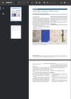
In this case study, a 15-year-old obese boy was diagnosed with acanthosis nigricans and hair casts in the axillae, with Wood's lamp and dermoscopic examination revealing specific features, yet fungal infection was ruled out through a negative KOH test.
15 citations
,
June 2020 in “Journal of the American Academy of Dermatology” Shiny white structures in trichoscopy can indicate long-standing discoid lupus erythematosus alopecia.
In a harbor seal from the Yellow Sea of Korea, this study found that alopecia-related lesions disappeared completely after treatment with internal and external ketoconazole, povidone iodine, cephalexin, and multivitamins over 8 months.
 4 citations
,
January 2016 in “Case reports in dermatological medicine”
4 citations
,
January 2016 in “Case reports in dermatological medicine” This case report describes a novel dermoscopic finding of blue-grey dots in a target pattern around yellow dots and follicles in radiation-induced alopecia following cerebral angiography with fluoroscopy.