 August 2026 in “Lasers in Medical Science”
 This review suggests that hyperspectral and terahertz imaging show promise in non-invasively assessing scalp changes associated with hair loss, but further validation is needed for clinical application.
 150 citations
,
April 2008 in “Journal of The American Academy of Dermatology”
150 citations
,
April 2008 in “Journal of The American Academy of Dermatology” This study found that videodermoscopy reveals distinct dermoscopic features in patients with alopecia areata incognita, aiding in biopsy decisions by identifying the appropriate sampling area and potentially avoiding unnecessary procedures.
12 citations
,
August 2001 in “PubMed” This study found that contrast enhanced phototrichogram (CE-PTG) significantly improved detection of various hair types and growth stages in androgenetic alopecia compared to standard PTG and was comparable to biopsy analysis.
2 citations
,
December 2018 This study found that dermatoscopy can quickly and non-invasively detect facial vellus hair involvement in patients with alopecia frontal fibrosante, occurring frequently and associated with specific facial lesions.
December 2020 in “Journal of Face Aesthetics” This study found that trichoscopy, a non-invasive diagnostic method, may effectively differentiate between types of focal alopecia in the parietal area in adult women, indicating specific trichoscopic features for conditions like alopecia areata, discoid lupus erythematosus, and lichen planopilaris.
 7 citations
,
August 2015 in “Dermatologic Surgery”
7 citations
,
August 2015 in “Dermatologic Surgery” This study found that cross beam laser is a reliable tool for assessing scalp laxity in hair restoration surgery, with results comparable to the Mayer elasticity scale.
1 citations
,
July 2025 in “Indian Dermatology Online Journal” This source outlines the crucial steps and techniques for conducting scalp biopsies to diagnose cicatricial and non-cicatricial alopecias, emphasizing the importance of proper biopsy site selection, technique, and specimen processing to accurately interpret histopathology and understand related hair follicle disorders.
 1 citations
,
October 2022 in “Asian journal of medical sciences”
1 citations
,
October 2022 in “Asian journal of medical sciences” This study found trichoscopy to be a highly sensitive, non-invasive method for diagnosing tinea capitis in children, which could be valuable in resource-limited settings lacking mycological culture facilities.
 1 citations
,
October 2023 in “Skin research and technology”
1 citations
,
October 2023 in “Skin research and technology” This study found that line-field confocal optical coherence tomography effectively visualized key diagnostic features of classic lichen planopilaris in real time, suggesting its potential as a valuable diagnostic tool.
17 citations
,
October 2001 in “British Journal of Ophthalmology” This report highlights a case where intralesional cidofovir successfully treated squamous cell carcinoma without systemic toxicity, suggesting it may be a viable alternative to surgical excision.
 50 citations
,
December 2011 in “Skin Research and Technology”
50 citations
,
December 2011 in “Skin Research and Technology” In this study, the researchers reported that their novel algorithm for hair restoration in dermoscopy images achieved high accuracy and texture preservation, outperforming other techniques in diagnostic accuracy and texture quality measures.
 70 citations
,
June 2003 in “Journal of Investigative Dermatology Symposium Proceedings”
70 citations
,
June 2003 in “Journal of Investigative Dermatology Symposium Proceedings” This study reports that the TrichoScan method effectively measures hair growth parameters and detected significant improvements in hair counts and thickness in men with androgenetic alopecia after finasteride treatment.
 February 2024 in “Skin research and technology”
 This study found that line-field confocal optical coherence tomography effectively visualized microscopic alterations in alopecia areata, suggesting its potential use in diagnosis and monitoring.
 10 citations
,
March 2014 in “International Journal of Dermatology”
10 citations
,
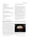
March 2014 in “International Journal of Dermatology” This report describes a case of postoperative pressure-induced alopecia in a 5-year-old girl following liver transplantation, suggesting that scalp dermatoscopy may help differentiate this condition from alopecia areata.

This study reviewed and reclassified 37 cases of primary scarring alopecia using histological methods to achieve more accurate diagnoses within subtypes like lupus erythematosus and lichen planopilaris.
 2 citations
,
May 2014 in “Journal of the Egyptian Women's Dermatologic Society (Print)”
2 citations
,
May 2014 in “Journal of the Egyptian Women's Dermatologic Society (Print)” This review examines the use of dermoscopy for diagnosing common hair disorders and highlights its importance, but it does not report new clinical results.
 December 2023 in “Portuguese journal of dermatology and venereology”
 This study found that trichoscopic examination can effectively differentiate between alopecia areata and trichotillomania, as specific features such as black dots and exclamation marks favor alopecia areata while hair breaking at various lengths and several unique patterns are exclusive to trichotillomania.
172 citations
,
December 1994 in “The Journal of Dermatologic Surgery and Oncology” This study introduces a hair transplantation method combining elliptical excision and micrografting, suggesting it provides significant cosmetic improvements for androgenetic alopecia.
 22 citations
,
May 2002 in “Skin Research and Technology”
22 citations
,
May 2002 in “Skin Research and Technology” This study found that in male androgenetic alopecia, the contrast enhanced phototrichogram technique was superior in detecting changes in hair density and thickness compared to other methods.
August 2003 in “Special report” This report introduces Barcelona - TrichoScan as a novel software tool purported to effectively analyze hair growth in patients with androgenetic alopecia or androgenetic effluvium, but it provides no clinical data.
December 2021 in “IP Indian journal of clinical and experimental dermatology” In this study, trichoscopy was identified as a useful non-invasive tool to aid in the diagnosis of female pattern hair loss, with peri pilar sign and hair diameter variability being common findings.
 7 citations
,
January 2012 in “International Journal of Trichology”
7 citations
,
January 2012 in “International Journal of Trichology” A man with Woolly Hair Syndrome had very curly, fragile hair, and doctors used a special scalp examination to diagnose him without invasive tests.
 June 2026 in “EXPERIMENTAL & CLINICAL MEDICINE GEORGIA”
 In this study, researchers found a positive correlation between trichoscopic features and histomorphological findings, supporting trichoscopy as a valuable tool for assessing androgenetic alopecia severity and monitoring treatment outcomes.
4 citations
,
April 2014 in “Lasers in Surgery and Medicine” This observational study reports that a TRASER device using specific wavelengths for vascular and follicular targets showed both clinical responses and histological changes, suggesting its potential as an effective vascular and hair removal tool.
 October 2024 in “Dermatology Practical & Conceptual”
 The researchers recommend considering porokeratosis as a possible diagnosis for scalp lesions since accurate identification is crucial for management and new treatments like topical cholesterol/lovastatin show promising potential for treating porokeratosis.
 40 citations
,
December 2014 in “Indian Journal of Dermatology”
40 citations
,
December 2014 in “Indian Journal of Dermatology” This study found that trichoscopy can effectively differentiate scalp psoriasis from seborrheic dermatitis by identifying specific trichoscopic patterns such as hidden hairs, signet ring vessels, and comma vessels.
45 citations
,
December 2004 in “Forensic Science International” This study reports that using laser microdissection to isolate telogen hair follicles significantly improves DNA extraction efficiency for STR typing, reducing contamination from keratin.
 5 citations
,
January 2018 in “Skin Research and Technology”
5 citations
,
January 2018 in “Skin Research and Technology” This letter compares automated digital image analysis (TrichoScan) with manual marking of hairs in male patients with androgenetic alopecia but reports no new results.
31 citations
,
October 2018 in “Journal of the European Academy of Dermatology and Venereology” In this study, trichoscopic examination found distinct hair characteristics for diagnosing eyebrow loss: alopecia areata is marked by features like exclamation mark hairs, while frontal fibrosing alopecia shows dystrophic hairs and distinctive regrowth patterns.
 24 citations
,
September 2018 in “Lasers in Surgery and Medicine”
24 citations
,
September 2018 in “Lasers in Surgery and Medicine” This study shows that multiphoton microscopy can non-invasively distinguish between scarring and non-scarring alopecia by identifying certain morphological features, but further research is needed to confirm its clinical utility.