Histopathology of Scarring and Nonscarring Hair Loss
September 2012
in “
Dermatologic Clinics
”
scarring hair loss nonscarring hair loss cicatricial alopecia non-cicatricial alopecia biopsy technique hair cycle lichen planopilaris chronic cutaneous lupus erythematosus androgenetic alopecia telogen effluvium hair miniaturization telogen hairs alopecia areata trichotillomania psoriatic alopecia histopathological characteristics follicular destruction scarring histological features biopsy lupus male pattern baldness female pattern baldness hair thinning hair pulling disorder psoriasis-related hair loss histopathology
TLDR The conclusion is that accurate diagnosis of different types of hair loss requires careful examination of tissue samples and understanding of clinical symptoms.
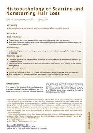
The 2013 document provides an in-depth analysis of the histopathological characteristics of scarring (cicatricial) and nonscarring (non-cicatricial) hair loss, emphasizing the importance of biopsy technique, hair cycle understanding, and clinical correlation for accurate diagnosis. It describes the optimal biopsy size and sectioning method, and differentiates between various hair loss conditions based on histological features. Cicatricial alopecias, such as lichen planopilaris and chronic cutaneous lupus erythematosus, show specific patterns of follicular destruction and scarring, while nonscarring alopecias, like androgenetic alopecia and telogen effluvium, are characterized by hair miniaturization and increased telogen hairs, respectively, without primary follicular destruction. The document also covers other hair loss disorders, including alopecia areata, trichotillomania, and psoriatic alopecia, detailing their unique histopathological findings. It stresses that despite overlapping features among different types of hair loss, accurate diagnosis can be achieved through careful histological examination combined with clinical information.
