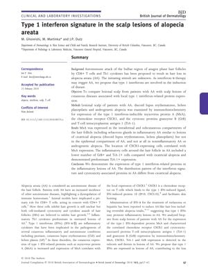
TLDR Alopecia areata shows a unique type 1 interferon signature, suggesting potential treatment by targeting this pathway.
The study examined the presence of type 1 interferon-related proteins in scalp lesions of patients with alopecia areata (AA) and compared them to other scalp conditions. Scalp biopsies from 6 patients with inflammatory AA, 2 with non-inflammatory AA, 2 with discoid lupus erythematosus (DLE), 2 with lichen planopilaris (LPP), and 2 with androgenetic alopecia were analyzed for the expression of myxovirus protein A (MxA), CXCR3, granzyme B (GrB), and T-cell intracytoplasmic antigen 1 (TiA-1). The results showed that MxA was expressed in inflammatory AA similar to cicatricial alopecia but not in non-inflammatory AA or androgenetic alopecia. CXCR3-expressing cells correlated with MxA expression, and AA lesions had fewer GrB+ cells and more TiA-1+ cells compared to cicatricial alopecia. The study concluded that AA features a type 1 interferon signature and differs from cicatricial alopecia in the pattern of interferon signature and cytotoxic proteins, suggesting AA is a cytotoxic assault on the hair follicle. Treatments that induce type 1 IFN were found ineffective for AA, indicating that targeting the IFN-α pathway may be a potential therapeutic approach.
 38 citations
,
January 2009 in “Journal of Cutaneous Medicine and Surgery”
 A woman developed hair loss after starting a treatment with adalimumab, suggesting this medication might cause hair loss.
 41 citations
,
August 2007 in “European Journal of Gastroenterology & Hepatology”
 A woman's total hair loss from hepatitis C treatment grew back after stopping the medication.
286 citations
,
August 2007 in “Journal of Clinical Investigation” Alopecia areata is an autoimmune disease where T cells attack hair follicles.
114 citations
,
August 2002 in “Journal of Investigative Dermatology” Alopecia areata is caused by an immune response, and targeting immune cells might help treat it.
 35 citations
,
January 2002 in “Dermatology”
 A woman's hair loss during treatment with specific hepatitis C drugs grew back after stopping the medication.
127 citations
,
January 2000 in “Journal of Investigative Dermatology” Cytotoxic T cells cause hair loss in chronic alopecia areata.
91 citations
,
March 1996 in “Archives of Dermatological Research” Certain cytokines and growth factors can inhibit hair growth and may affect alopecia areata.
 August 2018 in “Oxford University Press eBooks”
 The document's conclusion cannot be provided because the document cannot be parsed.
May 2018 in “Journal of cosmetology & trichology” Combining platelet-rich plasma therapy with prostaglandin-F eye drops can significantly regrow hair in alopecia universalis.
 36 citations
,
May 2011 in “Dermatologic therapy”
 No treatments fully cure or prevent alopecia areata; some help but have side effects or need more research.
