Trichoscopic Features of Frontal Fibrosing Alopecia: Results in 249 Patients
January 2015
in “
Journal of The American Academy of Dermatology
”
TLDR Trichoscopy helps diagnose and assess the severity of Frontal Fibrosing Alopecia.
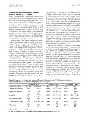
The study, conducted on 238 women diagnosed with Frontal Fibrosing Alopecia (FFA) across 12 Spanish centers, aimed to identify trichoscopic features of FFA and their correlation with disease severity and clinical parameters. Trichoscopic features such as cicatricial white patches, perifollicular erythema, follicular hyperkeratosis, lonely hairs, hair diameter diversity, and yellow dots were examined. The presence of cicatricial white patches was significantly associated with FFA severity, while perifollicular erythema and follicular hyperkeratosis were linked to pruritus. The study concluded that trichoscopy is useful for diagnosing FFA and assessing its activity, symptoms, and severity, despite limitations like its retrospective nature and evaluations being done by only two observers.
 nkrata