1 citations
,
January 2022 in “Skin Appendage Disorders” Lupus erythematosus can mimic alopecia areata, and trichoscopy is key for accurate diagnosis and better patient outcomes.
 9 citations
,
July 2020 in “Journal of Dermatology”
9 citations
,
July 2020 in “Journal of Dermatology” Asian patients with Frontal Fibrosing Alopecia often lose eyebrow hair and respond well to combined antiandrogen or antimalarial and topical treatments.
 1 citations
,
July 2020 in “Benha Journal of Applied Sciences”
1 citations
,
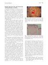
July 2020 in “Benha Journal of Applied Sciences” Trichoscopy is useful for diagnosing Frontal Fibrosing Alopecia.
15 citations
,
June 2020 in “Journal of the American Academy of Dermatology” Shiny white structures in trichoscopy can indicate long-standing discoid lupus erythematosus alopecia.
 9 citations
,
January 2020 in “Skin appendage disorders”
9 citations
,
January 2020 in “Skin appendage disorders” Hair loss from conditions like LPP and FFA can potentially be reversed with the right treatment.
 3 citations
,
August 2019 in “International Journal of Dermatology”
3 citations
,
August 2019 in “International Journal of Dermatology” Dermoscopy is useful for diagnosing lichen planopilaris and certain features may relate to disease duration, age, and gender.
 23 citations
,
November 2018 in “Journal of the European Academy of Dermatology and Venereology”
23 citations
,
November 2018 in “Journal of the European Academy of Dermatology and Venereology” The study concluded that severity of Frontal fibrosing alopecia is not linked to how long someone has it, can start before menopause, and eyebrow loss may be an early sign.
 9 citations
,
July 2018 in “International Journal of Dermatology”
9 citations
,
July 2018 in “International Journal of Dermatology” White and yellow dots indicate severe female hair loss in dark skin.
 12 citations
,
March 2018 in “Anais brasileiros de dermatologia/Anais Brasileiros de Dermatologia”
12 citations
,
March 2018 in “Anais brasileiros de dermatologia/Anais Brasileiros de Dermatologia” A patient had both chronic cutaneous lupus erythematosus and frontal fibrosing alopecia.
 34 citations
,
April 2016 in “International Journal of Dermatology”
34 citations
,
April 2016 in “International Journal of Dermatology” Trichoscopy is a useful method for identifying primary cicatricial alopecias and their specific types.
 38 citations
,
January 2016 in “Indian Journal of Dermatology, Venereology and Leprology”
38 citations
,
January 2016 in “Indian Journal of Dermatology, Venereology and Leprology” Trichoscopy is useful for diagnosing different types of hair loss.
 6 citations
,
January 2016 in “Skin appendage disorders”
6 citations
,
January 2016 in “Skin appendage disorders” A man with rare Lichen Planopilaris lost body hair, not scalp hair, and treatment stopped itching but didn't regrow hair.
 13 citations
,
July 2014 in “The Journal of Dermatology”
13 citations
,
July 2014 in “The Journal of Dermatology” Dermoscopy helped diagnose discoid lupus erythematosus in two patients without needing skin biopsies.
 28 citations
,
January 2014 in “Indian Journal of Dermatology, Venereology and Leprology”
28 citations
,
January 2014 in “Indian Journal of Dermatology, Venereology and Leprology” Chinese patients with primary cicatricial alopecia often have folliculitis decalvans, benefit from treatment, but may experience relapse, with dermoscopy being a useful diagnostic tool.
 34 citations
,
January 2014 in “International Journal of Trichology”
34 citations
,
January 2014 in “International Journal of Trichology” Polarized dermoscopy is slightly better than nonpolarized for diagnosing hair disorders, with each method having its own strengths.
 41 citations
,
January 2014 in “Annals of Dermatology”
41 citations
,
January 2014 in “Annals of Dermatology” Dermoscopic examination helps diagnose different types of hair loss conditions by showing specific patterns.
 96 citations
,
January 2013 in “International Journal of Trichology”
96 citations
,
January 2013 in “International Journal of Trichology” Trichoscopy is a useful, non-invasive way to diagnose different types of hair loss.
 62 citations
,
March 2012 in “Journal of the European Academy of Dermatology and Venereology”
62 citations
,
March 2012 in “Journal of the European Academy of Dermatology and Venereology” Using dermoscopy to guide scalp biopsies is an effective way to diagnose cicatricial alopecia.
 60 citations
,
September 2010 in “Journal of the American Academy of Dermatology”
60 citations
,
September 2010 in “Journal of the American Academy of Dermatology” Small white dots on the scalp seen with a dermoscope correspond to sweat ducts and vary with different hair disorders.
 73 citations
,
April 2010 in “Anais Brasileiros de Dermatologia”
73 citations
,
April 2010 in “Anais Brasileiros de Dermatologia” Dermoscopy helps diagnose and monitor treatment for hair loss from scarring conditions like discoid lupus and lichen planopilaris.
 33 citations
,
January 2010 in “International journal of trichology”
33 citations
,
January 2010 in “International journal of trichology” Antimicrobial therapy can help manage Folliculitis Decalvans.
 170 citations
,
December 2009 in “Histopathology”
170 citations
,
December 2009 in “Histopathology” The conclusion is that accurate diagnosis of different types of hair loss requires good teamwork between skin doctors and lab experts.
 61 citations
,
March 2009 in “The Journal of the American Board of Family Medicine”
61 citations
,
March 2009 in “The Journal of the American Board of Family Medicine” Early diagnosis and treatment of discoid lupus erythematosus improve outcomes.