Dermoscopy of Discoid Lupus Erythematosus: Report of Two Cases
July 2014
in “
The Journal of Dermatology
”
TLDR Dermoscopy helped diagnose discoid lupus erythematosus in two patients without needing skin biopsies.
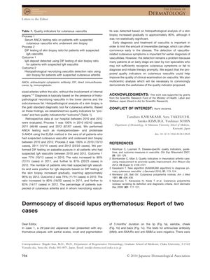
The document reports on two cases of discoid lupus erythematosus (DLE) and their dermoscopic findings. In the first case, a 29-year-old Japanese man presented with erythematous plaques on various parts of his body, and dermoscopy revealed features such as a follicular keratotic plug, polymorphous telangiectatic vessels, and white scales, among others. These findings, along with the clinical distribution and appearance of the plaques, led to a diagnosis of DLE, despite the patient's refusal of a skin biopsy. The second case involved a 39-year-old Japanese woman with a slightly erythematous plaque on her cheek. Dermoscopy showed follicular keratotic plugs and perifollicular pigmentation, and after treatment with oral prednisolone, her symptoms improved. Although a skin biopsy was not performed, she was diagnosed with DLE and lupus profundus based on dermoscopic findings and clinical course. The document also discusses the dermoscopic features of DLE in different phases of the disease and notes that perifollicular pigmentation, observed in the second case, has not been previously reported in DLE. The authors suggest that this report may be the first to describe perifollicular pigmentation in DLE.