T-Cell Reconstitution After Thymus Xenotransplantation Induces Hair Depigmentation and Loss
January 2013
in “
The journal of investigative dermatology/Journal of investigative dermatology
”
Studysummary This study found that thymus transplantation in athymic mice resulted in T-cell-driven hair follicle depigmentation and loss, without multiorgan autoimmune disease, highlighting mechanisms of tissue-specific tolerance. Our plain-language summary of this paper — not a Tressless recommendation.
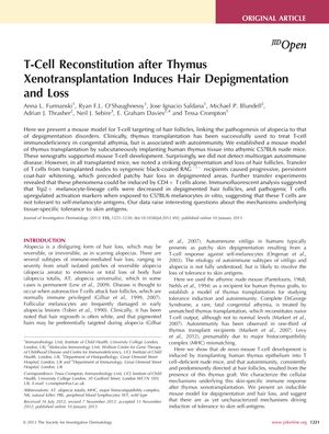
The study presented a mouse model demonstrating that T-cell reconstitution after thymus xenotransplantation could lead to hair depigmentation and loss, linking alopecia to depigmentation disorders. Human thymus tissue was implanted into athymic C57BL/6 nude mice, supporting mouse T-cell development without causing multiorgan autoimmune disease. However, all transplanted mice exhibited significant hair depigmentation and follicle loss. Transfer of T cells from these mice to black-coated RAG(-/-) recipients caused progressive coat-hair whitening and patchy hair loss. CD4+ T cells alone were sufficient to induce these effects. Immunofluorescent analysis indicated a decrease in Trp2+ melanocyte-lineage cells in depigmented follicles, and pathogenic T cells showed increased activation markers when exposed to melanocytes, suggesting a lack of tolerance to self-melanocyte antigens. This raised questions about tissue-specific tolerance mechanisms to skin antigens.