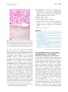
Horizontal and Vertical Sections of Scalp Biopsy Specimens from Dermatomyositis Patients with Scalp Involvement
February 2018
in “
Journal of The American Academy of Dermatology
”
scalp biopsy dermatomyositis atrophy mucinous stroma telangiectasia interface dermatitis follicular architecture sebaceous glands orthokeratosis hyperkeratosis reduced follicular density telogen count tortuous arborizing capillaries mucin deposition basement membrane thickening acrosyringeal hypergranulosis eosinophils chronic telogen effluvium non-scarring alopecia dermal mucin
TLDR Scalp biopsies from dermatomyositis patients show chronic hair loss without scarring, with mucin and blood vessel changes being very common.
The document provided a summary of a study examining scalp biopsy specimens from patients with dermatomyositis, revealing that the most common histopathologic findings were atrophy (90% of cases), mucinous stroma (100% of cases), telangiectasia in the papillary dermis (100% of cases), and interface dermatitis (85% of cases). The study also noted that follicular architecture was generally preserved, with sebaceous glands present in 81% of cases. Other findings included orthokeratosis and hyperkeratosis (95% of cases), reduced follicular density without miniaturization, normal telogen count, enlarged tortuous arborizing capillaries, moderate to significant mucin deposition (80% of cases), and basement membrane thickening (85% of cases). Unusual findings like acrosyringeal hypergranulosis with hyperkeratosis and the presence of eosinophils were also observed. The study concluded that the histopathologic features of scalp dermatomyositis are consistent with chronic telogen effluvium, with non-scarring alopecia, and that the presence of dermal mucin and telangiectasia were almost universal. Novel findings warranted further investigation. However, the number of participants was not provided, limiting the assessment of the study's strength.