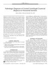
Pathologic Diagnosis of Central Centrifugal Cicatricial Alopecia on Horizontal Sections
September 2014
in “
American Journal of Dermatopathology
”
Central Centrifugal Cicatricial Alopecia CCCA follicular miniaturization inner root sheath desquamation sebaceous glands compound follicular structures perifollicular fibrosis perifollicular inflammation lamellar hyperkeratosis parakeratosis naked hair shafts goggles sign hair follicle shrinkage hair follicle structures fibrosis around hair follicles inflammation around hair follicles thickened skin layers skin cell retention exposed hair shafts
TLDR Horizontal sections of scalp biopsies are good for diagnosing Central Centrifugal Cicatricial Alopecia and help customize treatment.
The study analyzed scalp biopsies from 51 African-American women with Central Centrifugal Cicatricial Alopecia (CCCA) using horizontal sections to identify pathologic features. Key findings included follicular miniaturization in 81% of cases, premature desquamation of the inner root sheath in 96%, preservation of sebaceous glands in 94%, compound follicular structures with perifollicular fibrosis and/or inflammation in 89%, lamellar hyperkeratosis/parakeratosis in 79%, absent or mild inflammation in 77%, and naked hair shafts in 68%. The study concluded that horizontal sections are effective for diagnosing CCCA, especially in early or focal disease, and provide detailed information for tailoring treatment. It also suggested that hair breakage might trigger CCCA and highlighted the "goggles" sign and the preservation of sebaceous glands as important diagnostic features.