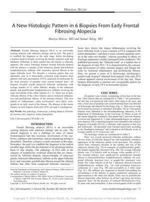
A New Histologic Pattern in 6 Biopsies From Early Frontal Fibrosing Alopecia
August 2018
in “
The American Journal of Dermatopathology
”
TLDR Researchers found a new early sign of Frontal Fibrosing Alopecia that could help avoid misdiagnosis.
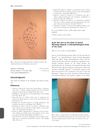
In the 2019 study involving 6 scalp biopsies from women with early Frontal Fibrosing Alopecia (FFA), researchers identified a new histologic pattern they termed "inflammatory vellus involvement." The biopsies, guided by trichoscopy, revealed preserved follicular architecture with an average of 11 vellus follicles, atrophied sebaceous glands, and perifollicular lymphohistiocytic infiltrate around the vellus follicles in 5 of the 6 cases, without perifollicular fibrosis or lichenoid inflammation. This pattern suggests an early stage of FFA and highlights the risk of misdiagnosis due to the absence of classic features in early FFA, underlining the need for awareness of this pattern to prevent delayed diagnosis and its associated impacts.

 nkrata