Distinguishing Histopathologic Features of Acantholytic Dermatoses and the Pattern of Acantholytic Hypergranulosis
September 2018
in “
Journal of cutaneous pathology
”

TLDR Different skin diseases show unique patterns of skin cell separation, cell death, and granular layer changes.
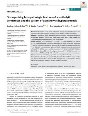
The retrospective review examined the histopathologic features of acantholysis in various skin diseases, including pemphigus vulgaris (PV), pemphigus foliaceus (PF), Hailey-Hailey disease (HHD), Darier disease (DD), Grover disease, and pityriasis rubra pilaris (PRP). The study included biopsies from 49 PV, 27 HHD, 25 DD, 65 Grover disease, 33 PRP, and 38 PF cases. It was found that adnexal acantholysis does not reliably distinguish PV from PF. Suprabasilar acantholysis was observed in PV, HHD, DD, Grover disease, and PRP, with PV and PRP showing acantholysis limited to the lower epidermis, while HHD, DD, and Grover disease involved all epidermal layers. PF mainly showed upper epidermal acantholysis. Follicular acantholysis was more frequent in PV and PF, and eccrine acantholysis was present in varying degrees across PV, HHD, PF, and DD. Grover disease, DD, and HHD had higher levels of dyskeratosis, and neutrophils were more common in PV, PF, and HHD, whereas eosinophils were more common in Grover disease and DD. Acantholytic hypergranulosis was predominantly seen in PF. The study concluded that the level of acantholysis, degree of dyskeratosis, and presence of acantholytic hypergranulosis are key features distinguishing between pemphigus types and other acantholytic disorders.
