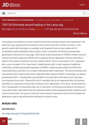
Full Thickness Wound Healing in the Lanyu Pig
April 2016
in “
Journal of Investigative Dermatology
”
TLDR Full thickness wounds on Lanyu pigs' skin resulted in abnormal skin structure and function due to changes in molecular expression patterns.
In 2016, researchers from the International Laboratory of Wound Repair and Regeneration at National Cheng Kung University in Taiwan used the Lanyu pig as an animal model to study full thickness wound healing due to its skin's similarity to human skin. They created 2x2cm, 2x3cm, 3x4cm, and 4x4cm full thickness wounds on the pigs' dorsum. By post wound day 57 (PWD57), they found no wound induced hair follicle neogenesis or papillary dermis regeneration. Wound contraction was greater in the anterior to posterior axis than medial to lateral. Healed wounds showed a decrease in K10+ suprabasal cells, an increase in K14+ basal cells, reduced p63 expression, less PCNA than unwounded tissue, more ColI and less ColIII, and failed to regenerate elastin protein. They also identified an alkaline phosphatase (ALP) + cell population in the basal layer at the base of each rete peg in normal pig and human skin that did not regenerate after full thickness wounding, hypothesizing that these cells contribute to skin regeneration. The study concluded that full thickness wounding on the dorsum of Lanyu pigs resulted in abnormal skin structure and function due to perturbed molecular expression patterns.