Characterization and Quantification of Wound-Induced Hair Follicle Neogenesis Using In Vivo Confocal Scanning Laser Microscopy
April 2011
in “
Skin Research and Technology
”
TLDR In vivo confocal scanning laser microscopy is an effective, non-invasive way to study and measure new hair growth after skin injury in mice.
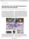
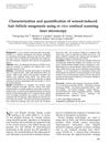
The study from 12 years ago demonstrated that in vivo confocal scanning laser microscopy (CSLM) is an effective and accurate method for non-invasively quantifying and characterizing neogenic hair follicles in mice after full-thickness wounds. CSLM detected 89% of hair follicles identified by K17 staining, outperforming alkaline phosphatase (AP) staining, and provided measurements that closely correlated with histological analysis. The study, which involved five mice, showed that CSLM could serially observe the development of hair follicles over time, offering a more dynamic and less invasive approach compared to traditional methods. CSLM was also found to avoid artifacts associated with histological processing and reduced the number of animals needed for research. The findings suggest that CSLM is a valuable tool for studying wound-induced hair follicle neogenesis and could be useful in evaluating treatment responses.