Yellow and Orange in Cutaneous Lesions: Clinical and Dermoscopic Data
September 2015
in “
Journal of the European Academy of Dermatology and Venereology
”
TLDR Yellow and orange colors are important for diagnosing certain skin conditions.
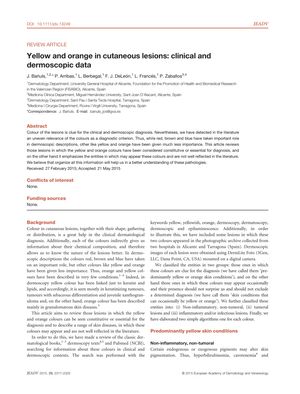
The document from 2015 reviews the significance of yellow and orange colors in diagnosing cutaneous lesions, noting that these colors have been less emphasized in literature compared to others. It classifies skin conditions into those predominantly yellow or orange and those that can occasionally exhibit these colors, including non-inflammatory, tumoral, and inflammatory or infectious lesions. Conditions such as hyperbilirubinemia, carotenemia, xanthomas, mastocytoma, sebaceous tumors, sarcoidosis, and leishmaniasis are discussed. Dermoscopic images from two hospitals in Spain illustrate the findings, and two diagnostic algorithms are provided. Yellow coloration is associated with conditions like alopecia areata, which can present with yellow dots in 54-75% of cases due to dilated infundibula of hair follicles. Orange hues are seen in conditions like targetoid haemosiderotic haemangioma and Spitz naevi. The presence of yellow in melanomas is rare but should not exclude the diagnosis. The document emphasizes the diagnostic importance of recognizing yellow and orange hues in skin diseases.

