Phenotypic Analysis of T-Cells in Extensive Alopecia Areata Scalp Suggests Partial Tolerance
TLDR T-cells in alopecia areata scalp show abnormal regulation, leading to less inflammation.
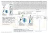
This study analyzed T-cell phenotypes and functional status in the scalp skin of 12 patients with extensive alopecia areata (EAA) and 6 control subjects using flow cytometry and immunohistochemical staining. EAA samples showed a lower percentage of CD-3-expressing cells, similar CD-4/CD-8 ratios to controls, and unique expression of CD-69, indicating T-cell activation. EAA T-cells produced less IL-2 and CD-8 T-cells produced less IFN-γ, with no increase in IFN-γ-producing cells compared to controls. These findings suggest that T-cell responses in EAA scalp are aberrantly regulated through peripheral T-cell tolerance mechanisms, potentially explaining the minimal inflammatory changes observed clinically.
View this study on jidonline.org →
Cited in this study
research The histopathology of alopecia areata in vertical and horizontal sections
Horizontal scalp biopsy sections are better for diagnosing alopecia areata, showing fewer hair follicles and more miniaturized hairs.
research Alopecia areata but not androgenetic alopecia is characterized by a restricted and oligoclonal T-cell receptor-repertoire among infiltrating lymphocytes
Alopecia areata involves specific T-cells, unlike androgenetic alopecia.
research Histopathology of non-scarring alopecia
The document describes how to tell different types of non-scarring hair loss apart by looking at hair and scalp tissue under a microscope.
research Alopecia areata: Pathogenesis and treatment
No single treatment is consistently effective for alopecia areata, and more research is needed.
Related
research Serum Zinc Concentration in Patients With Alopecia Areata
People with severe hair loss have lower zinc levels in their blood.