Characterizing The Expression Of Metabolic Markers In Alopecia Areata
alopecia areata hair follicle immunofluorescence metabolic transporters enzymes MPC1 Glyt1a xCT CD3+ cells SLC43A2 methionine transporter ASS1 arginine biosynthesis PCK2 pAMPK LDHA hair follicle mesenchyme immunometabolism T cells NK cells Liquid Chromatography-Mass Spectrometry LC-MS AA hair loss immune cells metabolic markers energy needs drug targets
TLDR The research identified unique metabolic activities in immune cells associated with hair loss in Alopecia Areata.
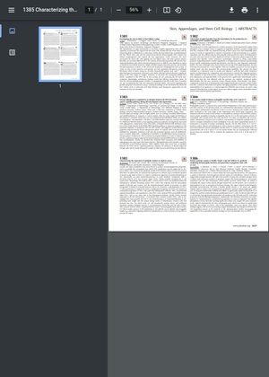
The study "Characterizing the expression of metabolic markers in alopecia areata" analyzed the metabolic profile of hair follicle immune cells in Alopecia Areata (AA). The researchers used immunofluorescence to mark metabolic transporters and enzymes in human scalp tissue sections. They found that the markers MPC1, Glyt1a, and xCT, which are involved in the uptake of glycine, cysteine, and pyruvate, were not expressed within the inflammatory infiltrate, including within CD3+ cells. However, a sub-population of CD3+ T cells co-expressed SLC43A2, a methionine transporter. ASS1, involved with arginine biosynthesis, was expressed in a few CD3+ cells, while PCK2 and pAMPK did not label CD3+ cells or any other cells in the inflammatory infiltrate. LDHA, broadly expressed in the hair follicle mesenchyme, co-localized with a limited number of CD3+ cells in AA. This research provides the first in situ characterization of immunometabolism in AA, offering insight into the unique energy needs of inflammatory immune cells that promote hair loss. Future work will quantitively analyze these and additional metabolic markers alongside specific T cell populations and NK cells, complemented by Liquid Chromatography-Mass Spectrometry (LC-MS), to identify new druggable targets to control immune cell function and prevent hair follicle attack.