29 citations
,
February 1989 in “Journal of Cutaneous Pathology” A unique hair tumor with a rippled pattern was identified, showing incomplete differentiation and unusual cell arrangements.
 1 citations
,
June 2023 in “Advances in therapy”
 Ripretinib is effective and safe for treating advanced GIST in Chinese patients, particularly for non-gastric GISTs.
 February 2025 in “International Journal of Molecular Sciences”
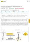
 RIPK1 inhibitors may help prevent alopecia areata by reducing immune cell activity.

RIPK1 inhibitors might help prevent alopecia areata.
September 2019 in “The journal of investigative dermatology/Journal of investigative dermatology” New RIPK4 gene mutations were found to cause a type of skin and limb birth defect.


