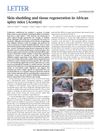
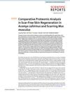
Comparative Transcriptomic Analysis of Dermal Wound Healing Reveals De Novo Skeletal Muscle Regeneration in Acomys Cahirinus
May 2019
in “
PLOS ONE
”
dermal wound healing skeletal muscle regeneration Acomys cahirinus Mus musculus panniculus carnosus myogenesis pathway myogenic regulatory factors embryonic myosin macrophage profiles inflammatory responses IL-10 anti-inflammatory cytokine Col6a1 satellite cell niche RNA-Seq RT-qPCR African spiny mouse common house mouse interleukin-10
TLDR The African spiny mouse can fully regenerate its muscle without scarring, unlike the common house mouse.
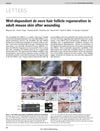
In the 2019 study by Brant et al., researchers compared the dermal wound healing process between the African spiny mouse (Acomys cahirinus) and the common house mouse (Mus musculus) through transcriptomic analysis. They found that Acomys cahirinus can regenerate skeletal muscle de novo, specifically the panniculus carnosus, after full-thickness excisional wounds without scarring, a process not observed in Mus musculus. This regeneration in Acomys cahirinus involves reactivation of the myogenesis pathway, upregulation of myogenic regulatory factors, and embryonic myosin, especially between days 14 and 18 post-wounding. The study also highlighted differences in macrophage profiles and inflammatory responses, with Acomys cahirinus showing higher levels of IL-10, an anti-inflammatory cytokine. Additionally, the upregulation of Col6a1, a component of the satellite cell niche, was noted in Acomys cahirinus. The research included RNA-Seq performed on 4 replicates per time point for each species and RT-qPCR on 3 replicates per time point for each species. This study is significant as it provides the first detailed analysis of de novo skeletal muscle regeneration in an adult mammal, which could have implications for regenerative medicine in humans.