Evaluation of Retinal Layers and Choroidal Structures Using Optical Coherence Tomography in Alopecia Areata
June 2023
in “
Medeniyet medical journal
”

TLDR People with alopecia areata have similar retinal structures but thicker choroidal regions compared to those without the condition.
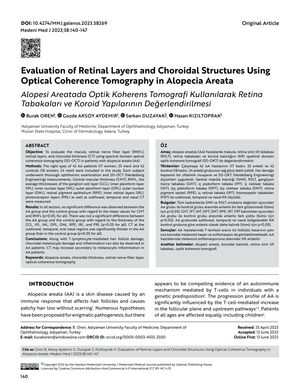
The study aimed to evaluate various retinal and choroidal structures in 42 patients with alopecia areata (AA) compared to 42 controls using spectral domain optical coherence tomography (SD-OCT). The measurements included central macular thickness (CMT), retinal nerve fiber layer (RNFL), and the thicknesses of several retinal layers, as well as choroidal thickness (CT) at different regions. The results showed no significant difference in the thickness of the CMT, RNFL, and retinal layers between the AA group and the control group. However, the CT was significantly thicker in the AA group in the subfoveal, temporal, and nasal regions. This suggests that along with T-lymphocyte-mediated hair follicle damage, AA patients may also experience choroidal melanocyte damage and inflammation, potentially leading to an increase in CT.


