Pilomatricoma: A Case Report and Intraoral Surgical Approach
September 2020
in “
Oral surgery, oral medicine, oral pathology and oral radiology
”
New to Keratin? There is a guide in the encyclopedia. Read the guide → Studysummary This case report describes a pilomatricoma in a 21-year-old woman, highlighting the diagnostic process and illustrating the potential for intraoral surgical access to minimize scarring.
Our plain-language summary. Not medical advice or a treatment recommendation. Consult a qualified healthcare professional before changing treatment. Full disclaimer
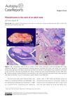
A 21-year-old white female presented with a 2 cm painless lesion in the right nasolabial fold, initially suspected to be a sebaceous cyst. An excisional biopsy via intraoral access was performed for esthetic reasons. Histopathologic examination revealed ghost cells, basaloid cells, keratin, calcifications, and chondroid material, leading to a diagnosis of pilomatricoma. This case highlighted the importance of oral pathologists being aware of skin lesions near the oral mucosa and demonstrated that intraoral surgical excision can avoid visible scarring.