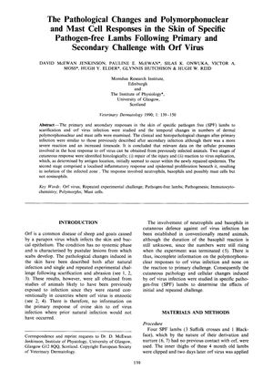
The Pathological Changes and Polymorphonuclear and Mast Cell Responses in the Skin of Specific Pathogen-Free Lambs Following Primary and Secondary Challenge with Orf Virus
September 1990
in “
Veterinary Dermatology
”
orf virus skin responses primary infection secondary infection clinical changes histopathological changes cellular immune response fibrocyte proliferation epidermal cell proliferation new epidermis neutrophils basophils mast cells degranulation erythema dendritic cells lymphocytes epithelial repair processes skin responses primary infection secondary infection clinical changes histopathological changes immune response fibrocyte proliferation epidermal cell proliferation new epidermis neutrophils basophils mast cells degranulation erythema dendritic cells lymphocytes epithelial repair

TLDR Lambs' skin showed similar but more severe responses to a second orf virus infection, involving immune cells and new skin formation.
In the 1990 study, researchers examined the skin responses of four specific pathogen-free lambs to primary and secondary challenges with orf virus. They found that the clinical and histopathological changes were similar in both primary and secondary infections, with the secondary infection eliciting a more severe and prolonged response. The defense mechanisms involved a cellular immune response, fibrocyte and epidermal cell proliferation, and the creation of a new epidermis to isolate the infected area. Neutrophils and basophils were clearly involved in the response, while eosinophils were not, as they remained within blood vessels and did not increase in number. Mast cell numbers remained unchanged, but their degranulation may contribute to the erythema observed. The study concluded that animals with previous infection can provide reliable data on cellular processes in the host defense reaction, and highlighted the need for further research on the dynamics of dendritic cells and lymphocytes, the interactions between cell types, and the control of epithelial repair processes.


