51 citations
,
November 1998 in “The journal of investigative dermatology/Journal of investigative dermatology” Beard cells, unlike scalp cells, produce growth factors in response to testosterone, which may explain differences in hair growth.
27 citations
,
December 1997 in “Archives of Dermatological Research” Rat dermal papilla cells have unique properties and interact differently with their environment compared to other skin cells.
11 citations
,
October 1997 in “British Journal of Dermatology” Wool follicles grew fibres for 8-10 days in a serum-free culture, influenced by calcium, glucose, amino acids, and insulin.
 19 citations
,
October 1996 in “Dermatologic Clinics”
19 citations
,
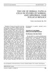
October 1996 in “Dermatologic Clinics” Dermal papilla cells are key for hair growth and could help us understand and treat hair loss.
36 citations
,
September 1996 in “PubMed” DP and DS cells are different from DF cells in structure and function.
37 citations
,
June 1996 in “Journal of cellular physiology” Retinoic acid, glucocorticoids, and IGF1 increase IGFBP-3 production in human dermal papilla cells, affecting hair growth.
9 citations
,
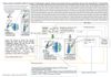
July 1993 in “The journal of investigative dermatology/Journal of investigative dermatology” Sex hormones and antiandrogens can either stimulate or inhibit human hair follicle cell growth depending on the dose.
34 citations
,
July 1993 in “The journal of investigative dermatology/Journal of investigative dermatology” Human hair growth is influenced by androgen hormones, and red deer mane follicles have similar hormone receptors.
44 citations
,
July 1993 in “Journal of Investigative Dermatology” 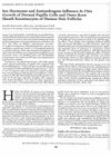 12 citations
,
July 1993 in “The journal of investigative dermatology/Journal of investigative dermatology”
12 citations
,
July 1993 in “The journal of investigative dermatology/Journal of investigative dermatology” Certain sex hormones and antiandrogens can either slow down or speed up the growth of human hair follicle cells depending on their concentration.
85 citations
,
January 1991 in “Journal of Investigative Dermatology” 22 citations
,
July 1990 in “Acta Dermato Venereologica” High levels of testosterone and dihydrotestosterone inhibit hair cell growth, while high levels of estradiol promote it.
106 citations
,
April 1986 in “British Journal of Dermatology” Dermal papilla cells from human hair follicles form unique structures and don't live as long as other skin cells in lab conditions.
 MagicBold