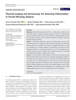
TLDR Infrared thermography, especially with dermoscopy, improves accuracy in diagnosing active hair loss due to inflammation.
The study, conducted on 22 female patients with Frontal Fibrosing Alopecia (FFA), evaluated the effectiveness of infrared thermography and optical dermoscopy in determining the activity of the inflammatory process in FFA. It found that thermography identified 77% of patients as having active FFA, which was more than the 64% identified through histological assessment. When combined with dermoscopy, the methods achieved a sensitivity of 64%, specificity of 88%, positive predictive value of 90%, and negative predictive value of 58% for diagnosing active FFA. The study concluded that infrared thermography, especially when used alongside dermoscopy, can enhance the accuracy of diagnosing the active phase of FFA, which is essential for appropriate treatment.
 95 citations
,
November 2016 in “Journal of The American Academy of Dermatology”
 Treatments for permanent hair loss from scarring aim to stop further loss, not regrow hair, and vary by condition, with partial success common.
 46 citations
,
January 2015 in “Journal of The American Academy of Dermatology”
 Trichoscopy helps diagnose and assess the severity of Frontal Fibrosing Alopecia.
 339 citations
,
February 2014 in “Journal of The American Academy of Dermatology”
 Most patients with frontal fibrosing alopecia are postmenopausal women, and treatments like finasteride and dutasteride can improve or stabilize the condition.
 62 citations
,
March 2012 in “Journal of the European Academy of Dermatology and Venereology”
 Using dermoscopy to guide scalp biopsies is an effective way to diagnose cicatricial alopecia.
159 citations
,
August 2010 in “British journal of dermatology/British journal of dermatology, Supplement” Hydroxychloroquine effectively reduces symptoms of frontal fibrosing alopecia, especially in the first 6 months.
 9 citations
,
January 2020 in “Postepy Dermatologii I Alergologii”
 Frontal fibrosing alopecia is a poorly understood condition with increasing cases and unclear treatment effectiveness.
 September 2017 in “Journal of Investigative Dermatology”
 HIF-1A may aid hair growth, Backhousia citriodora improves skin, autologous cells stabilize hair loss, infrared thermography assesses alopecia, and a new treatment preserves hair.
 September 2017 in “Journal of Investigative Dermatology”
 Thermal imaging is a useful non-invasive method to diagnose active inflammation in frontal fibrosing alopecia.
 September 2017 in “Journal of Investigative Dermatology”
 Injections of special skin cells showed potential in treating hair loss, with some participants experiencing increased hair density.
 1 citations
,
September 2017 in “Journal of Investigative Dermatology”
 Backhousia citriodora leaf extract effectively reduces oily skin across different ethnic groups.
