Lentiginous Melanoma: A Clinically Malignant Entity That Histopathologically Seems Benign. Case Study Harboring BRAF V600R Mutation
March 2015
in “
Journal of the European Academy of Dermatology and Venereology
”
TLDR A clinically suspected melanoma appeared benign under the microscope but was confirmed by specific tests and a rare mutation.
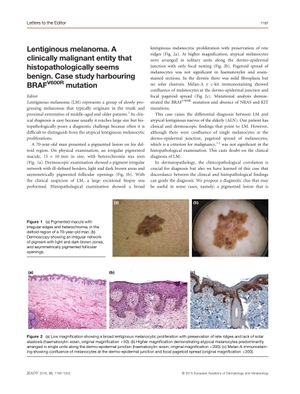
The document presents a case study of a 70-year-old man with a pigmented lesion on his deltoid region, which was clinically suspected to be lentiginous melanoma (LM), a type of melanoma that can appear benign histopathologically. Despite clinical and dermoscopic findings suggesting LM, the histopathological examination did not show significant pagetoid spread of melanocytes, a criterion for malignancy, leading to diagnostic uncertainty. Immunostaining with Melan-A and c-kit highlighted a confluent growth pattern and focal pagetoid spread, supporting the LM diagnosis. The lesion harbored a BRAF V600R mutation, which is rare but associated with low-grade melanoma and intermittent sun exposure. The mutation is also found in 70-80% of dysplastic nevi, indicating it should not be used alone for diagnosis when LM resembles an atypical lentiginous nevus. The document suggests that the presence of the BRAF V600R mutation may be a molecular marker for this melanoma subtype, as BRAF mutations are common in early invasive melanomas but rare in in situ melanomas. The case emphasizes the importance of clinicopathological correlation in dermatopathology and proposes that a clinically malignant lesion that appears benign histopathologically, especially when supported by specific immunostaining, may favor an LM diagnosis.
