Histopathological Examination of Hair Matrix Cysts
May 2020
in “
International journal of dermatology and venereology
”
TLDR Hair matrix cysts are rare skin nodules with unique features, often needing surgical removal.
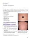
Hair matrix cysts (HMCs) were identified as rare hybrid cysts combining features of pilomatricoma and epidermal cysts, typically presenting as dark-red, dome-shaped nodules in children and adolescents. Histologically, HMCs exhibited a cyst wall with basaloid, transitional, and shadow cells, surrounded by a fibrous capsule with inflammatory cells. Key pathological features included basal-like cell layers, small cystic spaces, amorphous keratins, granulomatous reactions upon rupture, and beta-catenin accumulation indicating Wnt pathway activation. Differential diagnosis required histopathologic examination to distinguish HMCs from other cutaneous adnexal neoplasms. Treatment involved surgical resection and follow-up.