Histopathological and Dermoscopic Correlations in Pityriasis Versicolor
November 2022
in “
IP Indian journal of clinical and experimental dermatology
”
Pityriasis versicolor dermoscopic analysis ILLUCO Dermoscope pigmentary network scaling folliculocentric pattern contrast halo ring perivascular infiltrate hyperkeratosis hyphae spores spongiosis KOH mount yeast invasion of hair follicles PV dermoscope pigment network halo ring yeast in hair follicles
TLDR Dermoscopic and histopathological features combined can help diagnose Pityriasis versicolor.
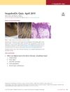
In a cross-sectional study involving 50 consecutive patients diagnosed with Pityriasis versicolor (PV), a correlation between dermoscopic and histopathological features was investigated. All patients tested positive for PV using KOH mount. Dermoscopic analysis using an ILLUCO Dermoscope revealed altered pigmentary network in almost all patients, scaling in 21 patients, a folliculocentric pattern in 20%, and a contrast halo ring in 4 patients. Histopathologically, perivascular infiltrate was observed in 78% of patients, hyperkeratosis in 62%, hyphae spores in 40%, and spongiosis in 42%. Special stains were used in 20 patients. The study concluded that combining dermoscopic features such as altered pigmentary network, contrast halo sign, and yeast invasion of hair follicles with histopathological features like perivascular infiltrate, hyperkeratosis, hyphae spores, and spongiosis can aid in the diagnosis of PV.