Fluoroscopy-Induced Morphea
January 2011
in “
Journal of The American Academy of Dermatology
”
TLDR A man developed a painful skin condition after multiple heart procedures involving radiation.
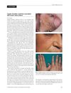
The document reports a case of fluoroscopy-induced morphea in a 64-year-old man who developed a painful and indurated plaque over his left scapula after undergoing six cardiac catheterizations. The plaque, which was erythematous, poikilodermatous, indurated, and atrophic, measured 9.0 × 6.0 cm. Biopsies revealed changes consistent with radiation-induced morphea, including epidermal acanthosis, hypermelanosis, increased collagen in a thickened sclerotic dermis, loss of follicular units, dilated vascular spaces, stellate fibroblasts, and a plasma cell infiltrate. Radiation-induced morphea is known to occur after exposure to radiation and is dependent on factors such as radiation dosage, beam projection, anatomic site, and duration of procedure. The condition has been previously reported in patients receiving radiation therapy for various cancers and those undergoing fluoroscopically guided procedures. Treatments for this condition have included corticosteroids, surgical excisions, and methotrexate with methylprednisolone, but due to underreporting, their effectiveness is not well-established.
