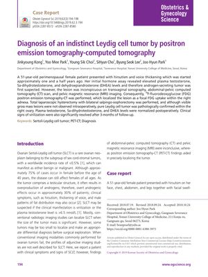
Diagnosis of an Indistinct Leydig Cell Tumor by Positron Emission Tomography-Computed Tomography
January 2019
in “
Obstetrics & gynecology science
”
Leydig cell tumor hirsutism voice thickening testosterone 5a-dihydrotestosterone DHEA androgen-secreting tumor 18F-fluorodeoxyglucose PET-CT total laparoscopic hysterectomy bilateral salpingo-oophorectomy right ovary virilization symptoms Leydig tumor hair growth deep voice DHT FDG PET-CT hysterectomy ovary removal male characteristics
TLDR A PET-CT scan successfully located a hard-to-find Leydig cell tumor in a woman with hormonal symptoms.
In 2019, a 51-year-old female patient with symptoms of hirsutism and voice thickening was diagnosed with a Leydig cell tumor, which was initially elusive to detection through transvaginal sonography, CT, and MRI. Elevated levels of testosterone, 5a-dihydrotestosterone, and DHEA suggested an androgen-secreting tumor, but it was only with the use of 18F-fluorodeoxyglucose PET-CT that the tumor was localized in the right adnexa. Following a total laparoscopic hysterectomy with bilateral salpingo-oophorectomy, the Leydig cell tumor was pathologically confirmed within the right ovary. The patient's hormone levels normalized, and the virilization symptoms significantly improved after a 3-month follow-up.
